sc-PDB
An Annotated Database of Druggable Binding Sites from the Protein DataBank
An Annotated Database of Druggable Binding Sites from the Protein DataBank
2.300 Å
X-ray
2002-09-16
| Name: | Ephrin type-A receptor 2 |
|---|---|
| ID: | EPHA2_HUMAN |
| AC: | P29317 |
| Organism: | Homo sapiens |
| Reign: | Eukaryota |
| TaxID: | 9606 |
| EC Number: | 2.7.10.1 |
| Chain Name: | Percentage of Residues within binding site |
|---|---|
| A | 100 % |
| B-Factor: | 41.264 |
|---|---|
| Number of residues: | 29 |
| Including | |
| Standard Amino Acids: | 29 |
| Non Standard Amino Acids: | 0 |
| Water Molecules: | 0 |
| Cofactors: | |
| Metals: | |
| Ligandability | Volume (Å3) |
|---|---|
| 0.923 | 610.875 |
| % Hydrophobic | % Polar |
|---|---|
| 48.07 | 51.93 |
| According to VolSite | |
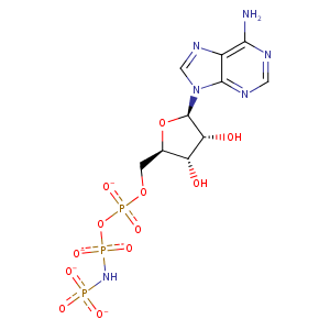
| HET Code: | ANP |
|---|---|
| Formula: | C10H13N6O12P3 |
| Molecular weight: | 502.164 g/mol |
| DrugBank ID: | - |
| Buried Surface Area: | 43.99 % |
| Polar Surface area: | 322.68 Å2 |
| Number of | |
|---|---|
| H-Bond Acceptors: | 16 |
| H-Bond Donors: | 4 |
| Rings: | 3 |
| Aromatic rings: | 2 |
| Anionic atoms: | 4 |
| Cationic atoms: | 0 |
| Rule of Five Violation: | 2 |
| Rotatable Bonds: | 8 |
| X | Y | Z |
|---|---|---|
| 38.3997 | 38.2485 | 49.3733 |
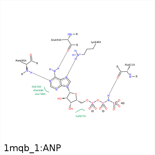


Represent the protein/ligand binding mode, centered on the ligand
Dashed lines represents hydrogen bonds and metal interactions
Green residue labels for amino acids with hydrophobic contacts (green lines) to the ligand
| Ligand | Protein | Interaction | |||
|---|---|---|---|---|---|
| Atom | Atom | Residue | Distance (Å) | Angle (°) | Type |
| C1' | CG1 | ILE- 619 | 3.88 | 0 | Hydrophobic |
| N3B | O | ALA- 621 | 3.08 | 148.9 | H-Bond (Ligand Donor) |
| C5' | CG2 | VAL- 627 | 4.25 | 0 | Hydrophobic |
| C1' | CG1 | VAL- 627 | 4.36 | 0 | Hydrophobic |
| O2A | NZ | LYS- 646 | 3.48 | 0 | Ionic (Protein Cationic) |
| N7 | NZ | LYS- 646 | 3.1 | 138.34 | H-Bond (Protein Donor) |
| N6 | O | GLU- 693 | 2.99 | 159.92 | H-Bond (Ligand Donor) |
| N1 | N | MET- 695 | 2.98 | 146.35 | H-Bond (Protein Donor) |
