sc-PDB
An Annotated Database of Druggable Binding Sites from the Protein DataBank
An Annotated Database of Druggable Binding Sites from the Protein DataBank
2.100 Å
X-ray
1998-03-27
| Name: | GTP cyclohydrolase 1 |
|---|---|
| ID: | GCH1_ECOLI |
| AC: | P0A6T5 |
| Organism: | Escherichia coli |
| Reign: | Bacteria |
| TaxID: | 83333 |
| EC Number: | 3.5.4.16 |
| Chain Name: | Percentage of Residues within binding site |
|---|---|
| B | 52 % |
| C | 48 % |
| B-Factor: | 30.156 |
|---|---|
| Number of residues: | 32 |
| Including | |
| Standard Amino Acids: | 29 |
| Non Standard Amino Acids: | 0 |
| Water Molecules: | 3 |
| Cofactors: | |
| Metals: | |
| Ligandability | Volume (Å3) |
|---|---|
| 0.351 | 418.500 |
| % Hydrophobic | % Polar |
|---|---|
| 44.35 | 55.65 |
| According to VolSite | |
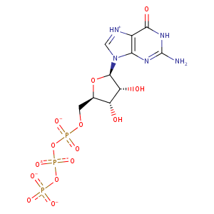
| HET Code: | GTP |
|---|---|
| Formula: | C10H12N5O14P3 |
| Molecular weight: | 519.149 g/mol |
| DrugBank ID: | DB04137 |
| Buried Surface Area: | 61.18 % |
| Polar Surface area: | 335.56 Å2 |
| Number of | |
|---|---|
| H-Bond Acceptors: | 17 |
| H-Bond Donors: | 4 |
| Rings: | 3 |
| Aromatic rings: | 1 |
| Anionic atoms: | 4 |
| Cationic atoms: | 0 |
| Rule of Five Violation: | 2 |
| Rotatable Bonds: | 8 |
| X | Y | Z |
|---|---|---|
| 165.455 | 23.8588 | 46.3366 |
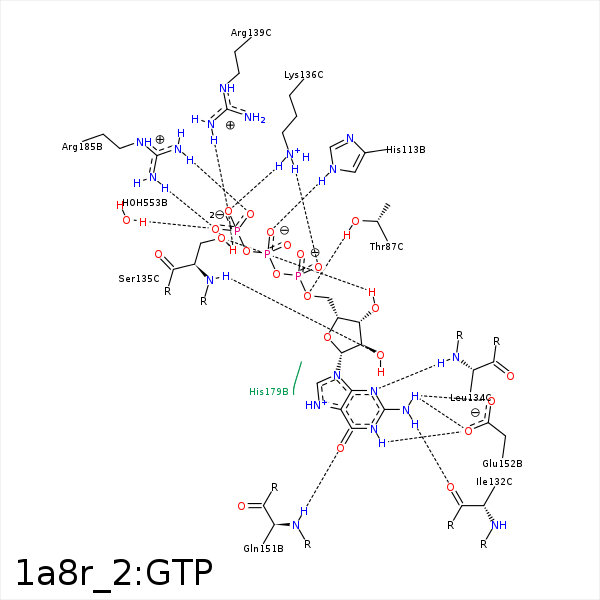


Represent the protein/ligand binding mode, centered on the ligand
Dashed lines represents hydrogen bonds and metal interactions
Green residue labels for amino acids with hydrophobic contacts (green lines) to the ligand
| Ligand | Protein | Interaction | |||
|---|---|---|---|---|---|
| Atom | Atom | Residue | Distance (Å) | Angle (°) | Type |
| O5' | OG1 | THR- 87 | 3.32 | 168.78 | H-Bond (Protein Donor) |
| C5' | CB | SER- 112 | 4.16 | 0 | Hydrophobic |
| O2B | NE2 | HIS- 113 | 2.79 | 147.54 | H-Bond (Protein Donor) |
| N2 | O | ILE- 132 | 3.06 | 149.77 | H-Bond (Ligand Donor) |
| N3 | N | LEU- 134 | 3.1 | 168.85 | H-Bond (Protein Donor) |
| O2G | OG | SER- 135 | 2.65 | 143.34 | H-Bond (Protein Donor) |
| O3G | OG | SER- 135 | 3.14 | 130.91 | H-Bond (Protein Donor) |
| O2' | N | SER- 135 | 2.98 | 152.24 | H-Bond (Protein Donor) |
| O3' | OG | SER- 135 | 2.73 | 157.52 | H-Bond (Ligand Donor) |
| C2' | CB | SER- 135 | 4.27 | 0 | Hydrophobic |
| O2G | NZ | LYS- 136 | 2.97 | 156.2 | H-Bond (Protein Donor) |
| O1A | NZ | LYS- 136 | 2.89 | 158.15 | H-Bond (Protein Donor) |
| O2G | NZ | LYS- 136 | 2.97 | 0 | Ionic (Protein Cationic) |
| O1B | NZ | LYS- 136 | 3.96 | 0 | Ionic (Protein Cationic) |
| O1A | NZ | LYS- 136 | 2.89 | 0 | Ionic (Protein Cationic) |
| O1G | CZ | ARG- 139 | 3.69 | 0 | Ionic (Protein Cationic) |
| O2G | CZ | ARG- 139 | 3.55 | 0 | Ionic (Protein Cationic) |
| O2G | NH1 | ARG- 139 | 2.64 | 132.27 | H-Bond (Protein Donor) |
| O6 | N | GLN- 151 | 2.82 | 166.51 | H-Bond (Protein Donor) |
| N1 | OE1 | GLU- 152 | 2.86 | 160.78 | H-Bond (Ligand Donor) |
| N2 | OE2 | GLU- 152 | 2.91 | 143.81 | H-Bond (Ligand Donor) |
| N2 | OE1 | GLU- 152 | 3.19 | 139.65 | H-Bond (Ligand Donor) |
| C2' | SG | CYS- 181 | 4.43 | 0 | Hydrophobic |
| O1G | NH2 | ARG- 185 | 2.87 | 153.85 | H-Bond (Protein Donor) |
| O3G | NH1 | ARG- 185 | 2.72 | 170.42 | H-Bond (Protein Donor) |
| O1G | CZ | ARG- 185 | 3.68 | 0 | Ionic (Protein Cationic) |
| O3G | CZ | ARG- 185 | 3.53 | 0 | Ionic (Protein Cationic) |
| O2B | CZ | ARG- 185 | 3.44 | 0 | Ionic (Protein Cationic) |
| O3G | O | HOH- 553 | 2.67 | 151.52 | H-Bond (Protein Donor) |
