sc-PDB
An Annotated Database of Druggable Binding Sites from the Protein DataBank
An Annotated Database of Druggable Binding Sites from the Protein DataBank
2.800 Å
X-ray
2015-05-02
| Name: | N-acetyl-beta-D glucosaminidase |
|---|---|
| ID: | V9M3A9_RHIMI |
| AC: | V9M3A9 |
| Organism: | Rhizomucor miehei |
| Reign: | Eukaryota |
| TaxID: | 4839 |
| EC Number: | / |
| Chain Name: | Percentage of Residues within binding site |
|---|---|
| B | 100 % |
| B-Factor: | 64.135 |
|---|---|
| Number of residues: | 46 |
| Including | |
| Standard Amino Acids: | 44 |
| Non Standard Amino Acids: | 0 |
| Water Molecules: | 2 |
| Cofactors: | |
| Metals: | |
| Ligandability | Volume (Å3) |
|---|---|
| 1.245 | 631.125 |
| % Hydrophobic | % Polar |
|---|---|
| 57.22 | 42.78 |
| According to VolSite | |
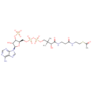
| HET Code: | ACO |
|---|---|
| Formula: | C23H34N7O17P3S |
| Molecular weight: | 805.539 g/mol |
| DrugBank ID: | - |
| Buried Surface Area: | 52.38 % |
| Polar Surface area: | 429.68 Å2 |
| Number of | |
|---|---|
| H-Bond Acceptors: | 22 |
| H-Bond Donors: | 5 |
| Rings: | 3 |
| Aromatic rings: | 2 |
| Anionic atoms: | 4 |
| Cationic atoms: | 0 |
| Rule of Five Violation: | 2 |
| Rotatable Bonds: | 20 |
| X | Y | Z |
|---|---|---|
| 107.783 | 177.026 | 150.436 |
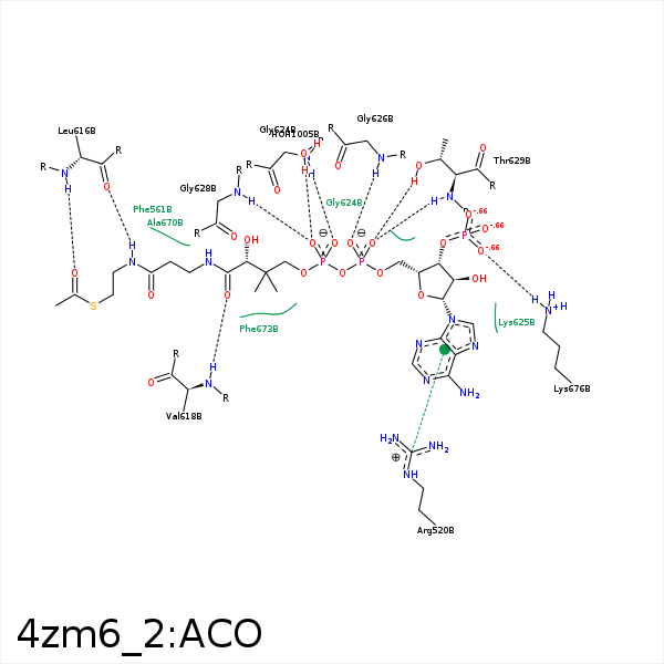


Represent the protein/ligand binding mode, centered on the ligand
Dashed lines represents hydrogen bonds and metal interactions
Green residue labels for amino acids with hydrophobic contacts (green lines) to the ligand
| Ligand | Protein | Interaction | |||
|---|---|---|---|---|---|
| Atom | Atom | Residue | Distance (Å) | Angle (°) | Type |
| N3A | NH2 | ARG- 520 | 3.33 | 120.62 | H-Bond (Protein Donor) |
| DuAr | CZ | ARG- 520 | 3.84 | 163 | Pi/Cation |
| C6P | CG2 | ILE- 560 | 3.86 | 0 | Hydrophobic |
| C6P | CZ | PHE- 561 | 3.81 | 0 | Hydrophobic |
| CDP | CD1 | LEU- 616 | 4.21 | 0 | Hydrophobic |
| N4P | O | LEU- 616 | 3.05 | 169.41 | H-Bond (Ligand Donor) |
| O | N | LEU- 616 | 2.94 | 137.71 | H-Bond (Protein Donor) |
| CDP | CG1 | VAL- 618 | 4.12 | 0 | Hydrophobic |
| O9P | N | VAL- 618 | 3.24 | 169.2 | H-Bond (Protein Donor) |
| CAP | CG | GLN- 623 | 3.93 | 0 | Hydrophobic |
| O5A | N | GLY- 624 | 3.16 | 147.98 | H-Bond (Protein Donor) |
| O1A | N | GLY- 626 | 2.83 | 151.75 | H-Bond (Protein Donor) |
| O4A | N | GLY- 628 | 2.9 | 152.97 | H-Bond (Protein Donor) |
| O2A | N | THR- 629 | 3.01 | 142.67 | H-Bond (Protein Donor) |
| O2A | OG1 | THR- 629 | 2.51 | 153.95 | H-Bond (Protein Donor) |
| S1P | CD2 | PHE- 657 | 4.26 | 0 | Hydrophobic |
| C2P | CG1 | VAL- 660 | 4.09 | 0 | Hydrophobic |
| CEP | CB | ALA- 670 | 3.76 | 0 | Hydrophobic |
| C2P | CB | ALA- 670 | 4.42 | 0 | Hydrophobic |
| C5B | CD2 | PHE- 673 | 4.49 | 0 | Hydrophobic |
| CCP | CE2 | PHE- 673 | 3.68 | 0 | Hydrophobic |
| CEP | CG | PHE- 673 | 4.17 | 0 | Hydrophobic |
| C2P | CE2 | PHE- 674 | 3.95 | 0 | Hydrophobic |
| O7A | NZ | LYS- 676 | 2.75 | 170.33 | H-Bond (Protein Donor) |
| O7A | NZ | LYS- 676 | 2.75 | 0 | Ionic (Protein Cationic) |
| O9A | NZ | LYS- 676 | 3.4 | 0 | Ionic (Protein Cationic) |
| O4A | O | HOH- 1005 | 2.76 | 165.12 | H-Bond (Protein Donor) |
