sc-PDB
An Annotated Database of Druggable Binding Sites from the Protein DataBank
An Annotated Database of Druggable Binding Sites from the Protein DataBank
2.300 Å
X-ray
2015-03-03
| Name: | D-amino-acid oxidase |
|---|---|
| ID: | OXDA_PIG |
| AC: | P00371 |
| Organism: | Sus scrofa |
| Reign: | Eukaryota |
| TaxID: | 9823 |
| EC Number: | 1.4.3.3 |
| Chain Name: | Percentage of Residues within binding site |
|---|---|
| B | 100 % |
| B-Factor: | 35.083 |
|---|---|
| Number of residues: | 71 |
| Including | |
| Standard Amino Acids: | 68 |
| Non Standard Amino Acids: | 0 |
| Water Molecules: | 3 |
| Cofactors: | |
| Metals: | |
| Ligandability | Volume (Å3) |
|---|---|
| 0.687 | 513.000 |
| % Hydrophobic | % Polar |
|---|---|
| 51.97 | 48.03 |
| According to VolSite | |
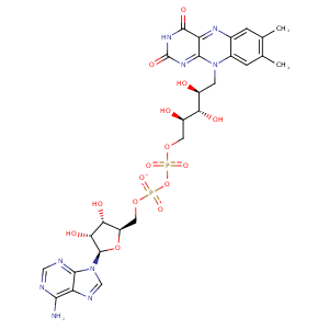
| HET Code: | FAD |
|---|---|
| Formula: | C27H31N9O15P2 |
| Molecular weight: | 783.534 g/mol |
| DrugBank ID: | DB03147 |
| Buried Surface Area: | 74.73 % |
| Polar Surface area: | 381.7 Å2 |
| Number of | |
|---|---|
| H-Bond Acceptors: | 22 |
| H-Bond Donors: | 7 |
| Rings: | 6 |
| Aromatic rings: | 3 |
| Anionic atoms: | 2 |
| Cationic atoms: | 0 |
| Rule of Five Violation: | 3 |
| Rotatable Bonds: | 13 |
| X | Y | Z |
|---|---|---|
| -33.4549 | -56.7924 | 11.2349 |


Represent the protein/ligand binding mode, centered on the ligand
Dashed lines represents hydrogen bonds and metal interactions
Green residue labels for amino acids with hydrophobic contacts (green lines) to the ligand
| Ligand | Protein | Interaction | |||
|---|---|---|---|---|---|
| Atom | Atom | Residue | Distance (Å) | Angle (°) | Type |
| O3B | N | ALA- 8 | 2.96 | 163.35 | H-Bond (Protein Donor) |
| C4' | CG1 | VAL- 10 | 4.25 | 0 | Hydrophobic |
| O2P | N | ILE- 11 | 3.29 | 157.45 | H-Bond (Protein Donor) |
| C1B | CB | ALA- 36 | 4.3 | 0 | Hydrophobic |
| O2B | OD1 | ASP- 37 | 2.52 | 167.71 | H-Bond (Ligand Donor) |
| N3A | N | ASP- 37 | 2.94 | 145.26 | H-Bond (Protein Donor) |
| O3B | O | ARG- 38 | 2.84 | 160.76 | H-Bond (Ligand Donor) |
| C2B | CG | ARG- 38 | 3.92 | 0 | Hydrophobic |
| O2B | N | ARG- 38 | 2.8 | 136.31 | H-Bond (Protein Donor) |
| C3B | CG2 | THR- 43 | 4.22 | 0 | Hydrophobic |
| O1A | OG1 | THR- 44 | 2.52 | 155.55 | H-Bond (Protein Donor) |
| C8M | CG2 | THR- 44 | 4.13 | 0 | Hydrophobic |
| O2A | N | THR- 45 | 3.06 | 149.39 | H-Bond (Protein Donor) |
| O4' | OG1 | THR- 45 | 2.96 | 159.32 | H-Bond (Ligand Donor) |
| C7M | CG1 | VAL- 47 | 4.12 | 0 | Hydrophobic |
| C6 | CB | ALA- 48 | 4.34 | 0 | Hydrophobic |
| C9A | CB | ALA- 48 | 4.01 | 0 | Hydrophobic |
| N5 | N | ALA- 49 | 2.98 | 161.35 | H-Bond (Protein Donor) |
| C6 | CB | ALA- 49 | 4.48 | 0 | Hydrophobic |
| O4 | N | GLY- 50 | 3.15 | 125.02 | H-Bond (Protein Donor) |
| N3 | O | LEU- 51 | 2.92 | 170.85 | H-Bond (Ligand Donor) |
| O4 | N | LEU- 51 | 3.2 | 171.95 | H-Bond (Protein Donor) |
| N6A | O | VAL- 164 | 2.83 | 176.54 | H-Bond (Ligand Donor) |
| N1A | N | VAL- 164 | 2.72 | 172.33 | H-Bond (Protein Donor) |
| C7M | CG2 | ILE- 202 | 3.85 | 0 | Hydrophobic |
| C5' | CG | PRO- 284 | 4.08 | 0 | Hydrophobic |
| O3' | O | GLY- 312 | 2.76 | 162.14 | H-Bond (Ligand Donor) |
| O3' | N | GLY- 315 | 3.13 | 128.66 | H-Bond (Protein Donor) |
| O2 | N | THR- 317 | 2.79 | 134.79 | H-Bond (Protein Donor) |
| O2 | OG1 | THR- 317 | 2.79 | 151.03 | H-Bond (Protein Donor) |
| O1P | O | HOH- 502 | 2.93 | 179.97 | H-Bond (Protein Donor) |
| O2P | O | HOH- 503 | 2.66 | 173.35 | H-Bond (Protein Donor) |
| O1P | O | HOH- 505 | 2.57 | 179.95 | H-Bond (Protein Donor) |
