sc-PDB
An Annotated Database of Druggable Binding Sites from the Protein DataBank
An Annotated Database of Druggable Binding Sites from the Protein DataBank
1.850 Å
X-ray
2014-12-05
| Name: | Dihydrofolate reductase |
|---|---|
| ID: | DYR_ECOLI |
| AC: | P0ABQ4 |
| Organism: | Escherichia coli |
| Reign: | Bacteria |
| TaxID: | 83333 |
| EC Number: | 1.5.1.3 |
| Chain Name: | Percentage of Residues within binding site |
|---|---|
| A | 100 % |
| B-Factor: | 18.559 |
|---|---|
| Number of residues: | 32 |
| Including | |
| Standard Amino Acids: | 30 |
| Non Standard Amino Acids: | 0 |
| Water Molecules: | 2 |
| Cofactors: | |
| Metals: | |
| Ligandability | Volume (Å3) |
|---|---|
| 0.935 | 891.000 |
| % Hydrophobic | % Polar |
|---|---|
| 41.29 | 58.71 |
| According to VolSite | |
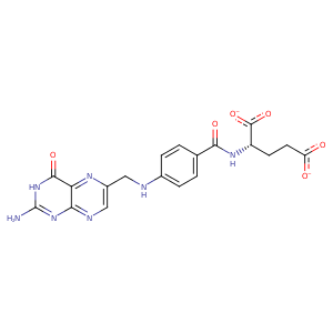
| HET Code: | FOL |
|---|---|
| Formula: | C19H17N7O6 |
| Molecular weight: | 439.382 g/mol |
| DrugBank ID: | DB00158 |
| Buried Surface Area: | 47.49 % |
| Polar Surface area: | 214.64 Å2 |
| Number of | |
|---|---|
| H-Bond Acceptors: | 12 |
| H-Bond Donors: | 4 |
| Rings: | 3 |
| Aromatic rings: | 2 |
| Anionic atoms: | 2 |
| Cationic atoms: | 0 |
| Rule of Five Violation: | 1 |
| Rotatable Bonds: | 9 |
| X | Y | Z |
|---|---|---|
| 21.4031 | 13.9165 | 13.9392 |
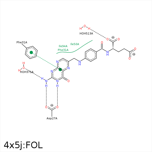


Represent the protein/ligand binding mode, centered on the ligand
Dashed lines represents hydrogen bonds and metal interactions
Green residue labels for amino acids with hydrophobic contacts (green lines) to the ligand
| Ligand | Protein | Interaction | |||
|---|---|---|---|---|---|
| Atom | Atom | Residue | Distance (Å) | Angle (°) | Type |
| NA2 | OD1 | ASP- 27 | 2.76 | 153.38 | H-Bond (Ligand Donor) |
| N3 | OD1 | ASP- 27 | 3.31 | 131.35 | H-Bond (Ligand Donor) |
| N3 | OD2 | ASP- 27 | 2.55 | 150.69 | H-Bond (Ligand Donor) |
| CB | CD | LYS- 32 | 4.26 | 0 | Hydrophobic |
| C9 | CG2 | THR- 46 | 3.78 | 0 | Hydrophobic |
| C15 | CD1 | ILE- 50 | 3.75 | 0 | Hydrophobic |
| C13 | CG1 | ILE- 50 | 3.49 | 0 | Hydrophobic |
| NA2 | O | HOH- 371 | 2.66 | 142.18 | H-Bond (Ligand Donor) |
| O1 | O | HOH- 513 | 2.68 | 179.97 | H-Bond (Protein Donor) |
