sc-PDB
An Annotated Database of Druggable Binding Sites from the Protein DataBank
An Annotated Database of Druggable Binding Sites from the Protein DataBank
2.000 Å
X-ray
2014-06-26
| Name: | Tubulin alpha-1B chain |
|---|---|
| ID: | TBA1B_BOVIN |
| AC: | P81947 |
| Organism: | Bos taurus |
| Reign: | Eukaryota |
| TaxID: | 9913 |
| EC Number: | / |
| Chain Name: | Percentage of Residues within binding site |
|---|---|
| A | 96 % |
| B | 4 % |
| B-Factor: | 46.575 |
|---|---|
| Number of residues: | 48 |
| Including | |
| Standard Amino Acids: | 45 |
| Non Standard Amino Acids: | 1 |
| Water Molecules: | 2 |
| Cofactors: | |
| Metals: | MG |
| Ligandability | Volume (Å3) |
|---|---|
| 0.288 | 884.250 |
| % Hydrophobic | % Polar |
|---|---|
| 32.06 | 67.94 |
| According to VolSite | |
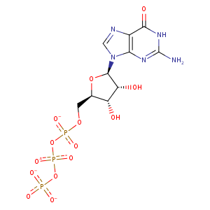
| HET Code: | GTP |
|---|---|
| Formula: | C10H12N5O14P3 |
| Molecular weight: | 519.149 g/mol |
| DrugBank ID: | DB04137 |
| Buried Surface Area: | 83.67 % |
| Polar Surface area: | 335.56 Å2 |
| Number of | |
|---|---|
| H-Bond Acceptors: | 17 |
| H-Bond Donors: | 4 |
| Rings: | 3 |
| Aromatic rings: | 1 |
| Anionic atoms: | 4 |
| Cationic atoms: | 0 |
| Rule of Five Violation: | 2 |
| Rotatable Bonds: | 8 |
| X | Y | Z |
|---|---|---|
| 22.3363 | 75.8573 | 50.2457 |


Represent the protein/ligand binding mode, centered on the ligand
Dashed lines represents hydrogen bonds and metal interactions
Green residue labels for amino acids with hydrophobic contacts (green lines) to the ligand
| Ligand | Protein | Interaction | |||
|---|---|---|---|---|---|
| Atom | Atom | Residue | Distance (Å) | Angle (°) | Type |
| O1B | N | GLN- 11 | 2.96 | 173.43 | H-Bond (Protein Donor) |
| N7 | NE2 | GLN- 11 | 3.3 | 145.36 | H-Bond (Protein Donor) |
| O2A | N | ALA- 12 | 3.09 | 165.04 | H-Bond (Protein Donor) |
| C1' | CB | ALA- 12 | 4.3 | 0 | Hydrophobic |
| O6 | NE2 | GLN- 15 | 2.66 | 142.91 | H-Bond (Protein Donor) |
| O1G | N | ALA- 99 | 2.81 | 156.69 | H-Bond (Protein Donor) |
| O3G | N | ASN- 101 | 2.79 | 150 | H-Bond (Protein Donor) |
| O2A | OG | SER- 140 | 2.91 | 153.09 | H-Bond (Protein Donor) |
| O5' | OG | SER- 140 | 3.12 | 134.75 | H-Bond (Protein Donor) |
| C4' | CB | SER- 140 | 3.96 | 0 | Hydrophobic |
| C1' | CB | SER- 140 | 4.41 | 0 | Hydrophobic |
| O3G | N | GLY- 144 | 2.93 | 146.42 | H-Bond (Protein Donor) |
| O1G | OG1 | THR- 145 | 2.57 | 144.57 | H-Bond (Protein Donor) |
| O3B | OG1 | THR- 145 | 3.37 | 124.13 | H-Bond (Protein Donor) |
| O3B | N | THR- 145 | 2.9 | 162.64 | H-Bond (Protein Donor) |
| O2B | N | GLY- 146 | 2.78 | 139.33 | H-Bond (Protein Donor) |
| C1' | CG2 | ILE- 171 | 4.49 | 0 | Hydrophobic |
| O3' | OE1 | GLU- 183 | 3.35 | 136.08 | H-Bond (Ligand Donor) |
| O3' | OE2 | GLU- 183 | 2.66 | 157.45 | H-Bond (Ligand Donor) |
| N2 | OD1 | ASN- 206 | 2.9 | 157.83 | H-Bond (Ligand Donor) |
| N3 | ND2 | ASN- 206 | 3.19 | 169.56 | H-Bond (Protein Donor) |
| C2' | CZ | TYR- 224 | 4.11 | 0 | Hydrophobic |
| O2' | OH | TYR- 224 | 3.08 | 153.2 | H-Bond (Protein Donor) |
| DuAr | DuAr | TYR- 224 | 3.87 | 0 | Aromatic Face/Face |
| O6 | ND2 | ASN- 228 | 2.92 | 163.22 | H-Bond (Protein Donor) |
| N1 | OD1 | ASN- 228 | 2.66 | 167.63 | H-Bond (Ligand Donor) |
| O2G | NZ | LYS- 254 | 2.88 | 171.87 | H-Bond (Protein Donor) |
| O2G | NZ | LYS- 254 | 2.88 | 0 | Ionic (Protein Cationic) |
| O3G | NZ | LYS- 254 | 3.95 | 0 | Ionic (Protein Cationic) |
| O2G | MG | MG- 502 | 1.98 | 0 | Metal Acceptor |
| O1B | MG | MG- 502 | 2.09 | 0 | Metal Acceptor |
| O3' | O | HOH- 611 | 2.79 | 179.95 | H-Bond (Protein Donor) |
