sc-PDB
An Annotated Database of Druggable Binding Sites from the Protein DataBank
An Annotated Database of Druggable Binding Sites from the Protein DataBank
2.240 Å
X-ray
2014-02-05
| Name: | Conserved Archaeal protein |
|---|---|
| ID: | Q4JA33_SULAC |
| AC: | Q4JA33 |
| Organism: | Sulfolobus acidocaldarius |
| Reign: | Archaea |
| TaxID: | 330779 |
| EC Number: | / |
| Chain Name: | Percentage of Residues within binding site |
|---|---|
| A | 100 % |
| B-Factor: | 46.192 |
|---|---|
| Number of residues: | 71 |
| Including | |
| Standard Amino Acids: | 67 |
| Non Standard Amino Acids: | 0 |
| Water Molecules: | 4 |
| Cofactors: | |
| Metals: | |
| Ligandability | Volume (Å3) |
|---|---|
| 0.432 | 546.750 |
| % Hydrophobic | % Polar |
|---|---|
| 37.04 | 62.96 |
| According to VolSite | |
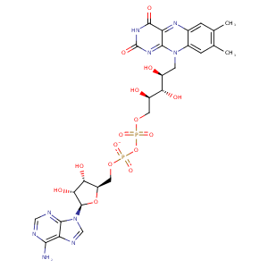
| HET Code: | FDA |
|---|---|
| Formula: | C27H33N9O15P2 |
| Molecular weight: | 785.550 g/mol |
| DrugBank ID: | - |
| Buried Surface Area: | 70.49 % |
| Polar Surface area: | 381.04 Å2 |
| Number of | |
|---|---|
| H-Bond Acceptors: | 21 |
| H-Bond Donors: | 9 |
| Rings: | 6 |
| Aromatic rings: | 3 |
| Anionic atoms: | 2 |
| Cationic atoms: | 0 |
| Rule of Five Violation: | 3 |
| Rotatable Bonds: | 13 |
| X | Y | Z |
|---|---|---|
| -11.943 | 23.5573 | -8.69843 |


Represent the protein/ligand binding mode, centered on the ligand
Dashed lines represents hydrogen bonds and metal interactions
Green residue labels for amino acids with hydrophobic contacts (green lines) to the ligand
| Ligand | Protein | Interaction | |||
|---|---|---|---|---|---|
| Atom | Atom | Residue | Distance (Å) | Angle (°) | Type |
| O1P | N | ALA- 16 | 3.21 | 165.35 | H-Bond (Protein Donor) |
| O3B | OD2 | ASP- 35 | 3.11 | 153.34 | H-Bond (Ligand Donor) |
| O2B | OD2 | ASP- 35 | 2.87 | 166.67 | H-Bond (Ligand Donor) |
| N3A | N | SER- 36 | 3.21 | 136.08 | H-Bond (Protein Donor) |
| O2' | NZ | LYS- 45 | 2.95 | 168.61 | H-Bond (Protein Donor) |
| C6 | CB | CYS- 47 | 4.45 | 0 | Hydrophobic |
| C9A | SG | CYS- 47 | 3.79 | 0 | Hydrophobic |
| C2' | SG | CYS- 47 | 4.04 | 0 | Hydrophobic |
| N5 | N | GLY- 48 | 2.99 | 145.47 | H-Bond (Protein Donor) |
| N3 | O | ALA- 50 | 2.88 | 137.25 | H-Bond (Ligand Donor) |
| O4 | N | ALA- 50 | 3.26 | 154.14 | H-Bond (Protein Donor) |
| N6A | O | ALA- 122 | 3.3 | 161.77 | H-Bond (Ligand Donor) |
| N1A | N | ALA- 122 | 3.06 | 157.5 | H-Bond (Protein Donor) |
| N7A | OG | SER- 162 | 3.09 | 133.34 | H-Bond (Protein Donor) |
| N6A | OG | SER- 162 | 3.11 | 156.37 | H-Bond (Ligand Donor) |
| C7M | CB | ALA- 185 | 3.59 | 0 | Hydrophobic |
| C7M | CG | ARG- 187 | 4.34 | 0 | Hydrophobic |
| C7M | CH2 | TRP- 217 | 4.1 | 0 | Hydrophobic |
| C8M | CB | ALA- 267 | 3.67 | 0 | Hydrophobic |
| C9 | CG2 | VAL- 269 | 3.49 | 0 | Hydrophobic |
| O3' | OD1 | ASP- 288 | 2.59 | 154.25 | H-Bond (Ligand Donor) |
| O3' | OD2 | ASP- 288 | 3.26 | 140.94 | H-Bond (Ligand Donor) |
| C5' | CB | ASP- 288 | 4.27 | 0 | Hydrophobic |
| O2P | N | ASP- 288 | 2.97 | 158.45 | H-Bond (Protein Donor) |
| N1 | N | GLY- 300 | 3.11 | 169.17 | H-Bond (Protein Donor) |
| O2 | N | GLY- 300 | 3.34 | 132.73 | H-Bond (Protein Donor) |
| O2 | N | LYS- 301 | 3 | 155.54 | H-Bond (Protein Donor) |
| O4' | NZ | LYS- 301 | 2.94 | 157.91 | H-Bond (Protein Donor) |
| C2' | CG | LYS- 301 | 4.18 | 0 | Hydrophobic |
| C4' | CG | LYS- 301 | 3.91 | 0 | Hydrophobic |
| O1A | O | HOH- 601 | 2.89 | 143.55 | H-Bond (Protein Donor) |
| O1P | O | HOH- 602 | 2.7 | 168.91 | H-Bond (Protein Donor) |
| O2P | O | HOH- 605 | 2.66 | 179.98 | H-Bond (Protein Donor) |
| O3B | O | HOH- 606 | 2.78 | 179.97 | H-Bond (Protein Donor) |
