sc-PDB
An Annotated Database of Druggable Binding Sites from the Protein DataBank
An Annotated Database of Druggable Binding Sites from the Protein DataBank
2.500 Å
X-ray
2013-02-08
| Name: | Thioredoxin reductase 2 |
|---|---|
| ID: | TRXR2_PLAF7 |
| AC: | P61076 |
| Organism: | Plasmodium falciparum |
| Reign: | Eukaryota |
| TaxID: | 36329 |
| EC Number: | 1.8.1.9 |
| Chain Name: | Percentage of Residues within binding site |
|---|---|
| A | 94 % |
| B | 6 % |
| B-Factor: | 35.075 |
|---|---|
| Number of residues: | 73 |
| Including | |
| Standard Amino Acids: | 69 |
| Non Standard Amino Acids: | 0 |
| Water Molecules: | 4 |
| Cofactors: | |
| Metals: | |
| Ligandability | Volume (Å3) |
|---|---|
| 1.175 | 1377.000 |
| % Hydrophobic | % Polar |
|---|---|
| 42.65 | 57.35 |
| According to VolSite | |
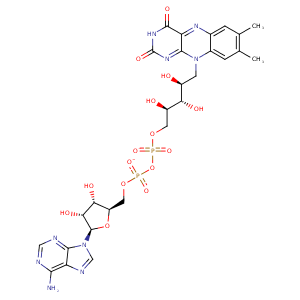
| HET Code: | FAD |
|---|---|
| Formula: | C27H31N9O15P2 |
| Molecular weight: | 783.534 g/mol |
| DrugBank ID: | DB03147 |
| Buried Surface Area: | 76.38 % |
| Polar Surface area: | 381.7 Å2 |
| Number of | |
|---|---|
| H-Bond Acceptors: | 22 |
| H-Bond Donors: | 7 |
| Rings: | 6 |
| Aromatic rings: | 3 |
| Anionic atoms: | 2 |
| Cationic atoms: | 0 |
| Rule of Five Violation: | 3 |
| Rotatable Bonds: | 13 |
| X | Y | Z |
|---|---|---|
| 23.07 | 49.7379 | 170.793 |


Represent the protein/ligand binding mode, centered on the ligand
Dashed lines represents hydrogen bonds and metal interactions
Green residue labels for amino acids with hydrophobic contacts (green lines) to the ligand
| Ligand | Protein | Interaction | |||
|---|---|---|---|---|---|
| Atom | Atom | Residue | Distance (Å) | Angle (°) | Type |
| C4' | CG | PRO- 51 | 4.26 | 0 | Hydrophobic |
| O1P | N | GLY- 52 | 2.77 | 152.84 | H-Bond (Protein Donor) |
| O3B | OD1 | ASP- 71 | 3.31 | 157.89 | H-Bond (Ligand Donor) |
| O2B | O | TYR- 72 | 3.14 | 162.68 | H-Bond (Ligand Donor) |
| N3A | N | TYR- 72 | 3.13 | 175.22 | H-Bond (Protein Donor) |
| O1A | OG1 | THR- 87 | 2.66 | 163.1 | H-Bond (Protein Donor) |
| O2A | N | THR- 87 | 3.01 | 168.27 | H-Bond (Protein Donor) |
| C8M | CG2 | THR- 87 | 3.71 | 0 | Hydrophobic |
| C2' | CB | CYS- 88 | 4.35 | 0 | Hydrophobic |
| C9A | SG | CYS- 93 | 4.36 | 0 | Hydrophobic |
| C2' | SG | CYS- 93 | 4.3 | 0 | Hydrophobic |
| O4 | NZ | LYS- 96 | 2.81 | 129.52 | H-Bond (Protein Donor) |
| N5 | NZ | LYS- 96 | 2.93 | 121.27 | H-Bond (Protein Donor) |
| C6 | CB | LYS- 96 | 4.4 | 0 | Hydrophobic |
| N6A | O | ALA- 161 | 3.01 | 156.03 | H-Bond (Ligand Donor) |
| N1A | N | ALA- 161 | 3.34 | 155.29 | H-Bond (Protein Donor) |
| C7M | CB | SER- 212 | 3.83 | 0 | Hydrophobic |
| C7M | CE2 | PHE- 216 | 4.07 | 0 | Hydrophobic |
| C7M | CG1 | VAL- 233 | 3.97 | 0 | Hydrophobic |
| C7 | CG2 | VAL- 233 | 4.14 | 0 | Hydrophobic |
| C8M | CD | ARG- 316 | 4.05 | 0 | Hydrophobic |
| O3' | OD1 | ASP- 357 | 3.08 | 169.56 | H-Bond (Ligand Donor) |
| C5' | CB | ASP- 357 | 4.15 | 0 | Hydrophobic |
| O2P | N | ASP- 357 | 2.83 | 165.07 | H-Bond (Protein Donor) |
| O2 | N | ALA- 366 | 3.12 | 136.34 | H-Bond (Protein Donor) |
| C4' | CB | ALA- 366 | 4.38 | 0 | Hydrophobic |
| C5' | CB | ALA- 369 | 4.35 | 0 | Hydrophobic |
| N3 | O | HIS- 509 | 2.72 | 148.51 | H-Bond (Ligand Donor) |
| O2P | O | HOH- 701 | 2.9 | 179.95 | H-Bond (Protein Donor) |
| O1A | O | HOH- 704 | 2.53 | 179.96 | H-Bond (Protein Donor) |
| O1P | O | HOH- 711 | 2.71 | 175.59 | H-Bond (Protein Donor) |
