sc-PDB
An Annotated Database of Druggable Binding Sites from the Protein DataBank
An Annotated Database of Druggable Binding Sites from the Protein DataBank
1.350 Å
X-ray
2012-06-04
| Name: | Ribosyldihydronicotinamide dehydrogenase [quinone] |
|---|---|
| ID: | NQO2_HUMAN |
| AC: | P16083 |
| Organism: | Homo sapiens |
| Reign: | Eukaryota |
| TaxID: | 9606 |
| EC Number: | / |
| Chain Name: | Percentage of Residues within binding site |
|---|---|
| A | 50 % |
| B | 50 % |
| B-Factor: | 13.004 |
|---|---|
| Number of residues: | 30 |
| Including | |
| Standard Amino Acids: | 27 |
| Non Standard Amino Acids: | 1 |
| Water Molecules: | 2 |
| Cofactors: | FAD |
| Metals: | |
| Ligandability | Volume (Å3) |
|---|---|
| 0.914 | 506.250 |
| % Hydrophobic | % Polar |
|---|---|
| 52.67 | 47.33 |
| According to VolSite | |
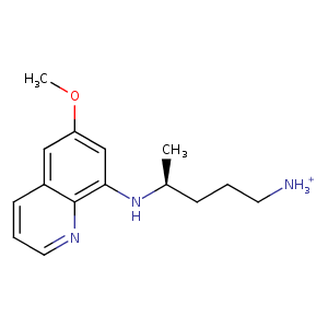
| HET Code: | 1PQ |
|---|---|
| Formula: | C15H22N3O |
| Molecular weight: | 260.355 g/mol |
| DrugBank ID: | - |
| Buried Surface Area: | 61.63 % |
| Polar Surface area: | 61.79 Å2 |
| Number of | |
|---|---|
| H-Bond Acceptors: | 3 |
| H-Bond Donors: | 2 |
| Rings: | 2 |
| Aromatic rings: | 2 |
| Anionic atoms: | 0 |
| Cationic atoms: | 1 |
| Rule of Five Violation: | 0 |
| Rotatable Bonds: | 6 |
| X | Y | Z |
|---|---|---|
| 23.5874 | -16.3605 | -14.7199 |
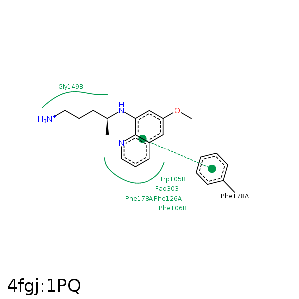


Represent the protein/ligand binding mode, centered on the ligand
Dashed lines represents hydrogen bonds and metal interactions
Green residue labels for amino acids with hydrophobic contacts (green lines) to the ligand
| Ligand | Protein | Interaction | |||
|---|---|---|---|---|---|
| Atom | Atom | Residue | Distance (Å) | Angle (°) | Type |
| C1 | CH2 | TRP- 105 | 3.47 | 0 | Hydrophobic |
| C1 | CZ | PHE- 126 | 3.32 | 0 | Hydrophobic |
| DuAr | DuAr | PHE- 178 | 3.56 | 0 | Aromatic Face/Face |
| DuAr | DuAr | PHE- 178 | 3.56 | 0 | Aromatic Face/Face |
| C13 | CB | PHE- 178 | 4.32 | 0 | Hydrophobic |
| N2 | OE1 | GLU- 193 | 3.36 | 0 | Ionic (Ligand Cationic) |
| N2 | OE2 | GLU- 193 | 3.3 | 0 | Ionic (Ligand Cationic) |
| C1 | C6 | FAD- 303 | 3.52 | 0 | Hydrophobic |
| C3 | C1' | FAD- 303 | 4.26 | 0 | Hydrophobic |
| C7 | C1' | FAD- 303 | 3.9 | 0 | Hydrophobic |
