sc-PDB
An Annotated Database of Druggable Binding Sites from the Protein DataBank
An Annotated Database of Druggable Binding Sites from the Protein DataBank
1.800 Å
X-ray
2012-04-19
| Name: | Coenzyme A disulfide reductase |
|---|---|
| ID: | CDR_STAA3 |
| AC: | Q2FIA5 |
| Organism: | Staphylococcus aureus |
| Reign: | Bacteria |
| TaxID: | 367830 |
| EC Number: | / |
| Chain Name: | Percentage of Residues within binding site |
|---|---|
| A | 91 % |
| B | 9 % |
| B-Factor: | 18.580 |
|---|---|
| Number of residues: | 64 |
| Including | |
| Standard Amino Acids: | 57 |
| Non Standard Amino Acids: | 1 |
| Water Molecules: | 6 |
| Cofactors: | COA |
| Metals: | |
| Ligandability | Volume (Å3) |
|---|---|
| 0.999 | 783.000 |
| % Hydrophobic | % Polar |
|---|---|
| 56.03 | 43.97 |
| According to VolSite | |
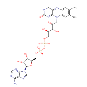
| HET Code: | FAD |
|---|---|
| Formula: | C27H31N9O15P2 |
| Molecular weight: | 783.534 g/mol |
| DrugBank ID: | DB03147 |
| Buried Surface Area: | 78.91 % |
| Polar Surface area: | 381.7 Å2 |
| Number of | |
|---|---|
| H-Bond Acceptors: | 22 |
| H-Bond Donors: | 7 |
| Rings: | 6 |
| Aromatic rings: | 3 |
| Anionic atoms: | 2 |
| Cationic atoms: | 0 |
| Rule of Five Violation: | 3 |
| Rotatable Bonds: | 13 |
| X | Y | Z |
|---|---|---|
| 23.4758 | 10.8416 | -11.8958 |


Represent the protein/ligand binding mode, centered on the ligand
Dashed lines represents hydrogen bonds and metal interactions
Green residue labels for amino acids with hydrophobic contacts (green lines) to the ligand
| Ligand | Protein | Interaction | |||
|---|---|---|---|---|---|
| Atom | Atom | Residue | Distance (Å) | Angle (°) | Type |
| C4B | CG2 | VAL- 10 | 4.14 | 0 | Hydrophobic |
| O2A | N | ALA- 11 | 3.27 | 159.85 | H-Bond (Protein Donor) |
| C4' | CB | ALA- 11 | 3.6 | 0 | Hydrophobic |
| O1P | N | GLY- 12 | 2.82 | 148.37 | H-Bond (Protein Donor) |
| O3B | OE1 | GLU- 33 | 2.69 | 171 | H-Bond (Ligand Donor) |
| O3B | OE2 | GLU- 33 | 3.1 | 125.93 | H-Bond (Ligand Donor) |
| O2B | OE2 | GLU- 33 | 2.86 | 173.08 | H-Bond (Ligand Donor) |
| N3A | N | LYS- 34 | 3.14 | 141 | H-Bond (Protein Donor) |
| C2B | CE | LYS- 34 | 3.89 | 0 | Hydrophobic |
| O2A | ND2 | ASN- 42 | 2.93 | 139.82 | H-Bond (Protein Donor) |
| C9 | CB | ASN- 42 | 4.12 | 0 | Hydrophobic |
| C9A | SG | CYS- 43 | 3.87 | 0 | Hydrophobic |
| C7M | CB | LEU- 45 | 4.26 | 0 | Hydrophobic |
| C6 | CG | PRO- 46 | 4.27 | 0 | Hydrophobic |
| C7M | CG | PRO- 46 | 3.93 | 0 | Hydrophobic |
| N6A | O | VAL- 81 | 2.84 | 168.74 | H-Bond (Ligand Donor) |
| N1A | N | VAL- 81 | 2.91 | 156.38 | H-Bond (Protein Donor) |
| O1P | OG | SER- 112 | 2.66 | 147.25 | H-Bond (Protein Donor) |
| C7M | CD2 | LEU- 130 | 3.64 | 0 | Hydrophobic |
| C8M | CD | ARG- 131 | 3.91 | 0 | Hydrophobic |
| N1 | OH | TYR- 158 | 3.43 | 127.92 | H-Bond (Protein Donor) |
| C6 | CG2 | VAL- 159 | 3.77 | 0 | Hydrophobic |
| C9A | CG2 | VAL- 159 | 4.42 | 0 | Hydrophobic |
| C7M | CG1 | VAL- 159 | 3.36 | 0 | Hydrophobic |
| O3' | OD1 | ASP- 277 | 2.85 | 177.43 | H-Bond (Ligand Donor) |
| O2P | N | ASP- 277 | 2.87 | 154.25 | H-Bond (Protein Donor) |
| N1 | N | ALA- 295 | 3.45 | 134.08 | H-Bond (Protein Donor) |
| O2 | N | ALA- 295 | 2.72 | 165.22 | H-Bond (Protein Donor) |
| C5' | CB | ALA- 298 | 4 | 0 | Hydrophobic |
| N3 | O | TYR- 419 | 2.96 | 171.73 | H-Bond (Ligand Donor) |
| O5B | O | HOH- 601 | 3.24 | 149.2 | H-Bond (Protein Donor) |
| O2P | O | HOH- 604 | 2.73 | 179.95 | H-Bond (Protein Donor) |
| O3B | O | HOH- 610 | 2.75 | 179.97 | H-Bond (Protein Donor) |
| O1A | O | HOH- 613 | 2.9 | 141.24 | H-Bond (Protein Donor) |
