sc-PDB
An Annotated Database of Druggable Binding Sites from the Protein DataBank
An Annotated Database of Druggable Binding Sites from the Protein DataBank
2.400 Å
X-ray
2012-04-12
| Name: | Toluene 1,2-dioxygenase system ferredoxin--NAD(+) reductase component |
|---|---|
| ID: | TODA_PSEP1 |
| AC: | A5W4E9 |
| Organism: | Pseudomonas putida |
| Reign: | Bacteria |
| TaxID: | 351746 |
| EC Number: | 1.18.1.3 |
| Chain Name: | Percentage of Residues within binding site |
|---|---|
| A | 100 % |
| B-Factor: | 12.327 |
|---|---|
| Number of residues: | 60 |
| Including | |
| Standard Amino Acids: | 55 |
| Non Standard Amino Acids: | 0 |
| Water Molecules: | 5 |
| Cofactors: | |
| Metals: | |
| Ligandability | Volume (Å3) |
|---|---|
| 1.232 | 1360.125 |
| % Hydrophobic | % Polar |
|---|---|
| 41.69 | 58.31 |
| According to VolSite | |
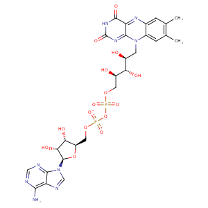
| HET Code: | FAD |
|---|---|
| Formula: | C27H31N9O15P2 |
| Molecular weight: | 783.534 g/mol |
| DrugBank ID: | DB03147 |
| Buried Surface Area: | 74.06 % |
| Polar Surface area: | 381.7 Å2 |
| Number of | |
|---|---|
| H-Bond Acceptors: | 22 |
| H-Bond Donors: | 7 |
| Rings: | 6 |
| Aromatic rings: | 3 |
| Anionic atoms: | 2 |
| Cationic atoms: | 0 |
| Rule of Five Violation: | 3 |
| Rotatable Bonds: | 13 |
| X | Y | Z |
|---|---|---|
| 31.4275 | 6.87662 | -8.14674 |


Represent the protein/ligand binding mode, centered on the ligand
Dashed lines represents hydrogen bonds and metal interactions
Green residue labels for amino acids with hydrophobic contacts (green lines) to the ligand
| Ligand | Protein | Interaction | |||
|---|---|---|---|---|---|
| Atom | Atom | Residue | Distance (Å) | Angle (°) | Type |
| C4' | CG1 | VAL- 12 | 4.14 | 0 | Hydrophobic |
| O1P | N | GLY- 13 | 2.99 | 138.14 | H-Bond (Protein Donor) |
| O2B | OD1 | ASP- 35 | 2.79 | 152.92 | H-Bond (Ligand Donor) |
| O1A | NE | ARG- 43 | 3.09 | 151.21 | H-Bond (Protein Donor) |
| O1A | CZ | ARG- 43 | 3.77 | 0 | Ionic (Protein Cationic) |
| O2A | CZ | ARG- 43 | 3.87 | 0 | Ionic (Protein Cationic) |
| C8 | CG | ARG- 43 | 4.11 | 0 | Hydrophobic |
| C9A | CG | PRO- 44 | 4.48 | 0 | Hydrophobic |
| C7M | CB | LEU- 46 | 4.29 | 0 | Hydrophobic |
| C7M | CB | SER- 47 | 3.82 | 0 | Hydrophobic |
| O4 | NZ | LYS- 48 | 3.19 | 146 | H-Bond (Protein Donor) |
| N5 | NZ | LYS- 48 | 3.35 | 135.65 | H-Bond (Protein Donor) |
| N6A | O | VAL- 80 | 3.02 | 162.92 | H-Bond (Ligand Donor) |
| N1A | N | VAL- 80 | 3.01 | 168.83 | H-Bond (Protein Donor) |
| C7M | CD2 | LEU- 127 | 4.23 | 0 | Hydrophobic |
| O2A | NH2 | ARG- 128 | 2.77 | 148.62 | H-Bond (Protein Donor) |
| O2A | CZ | ARG- 128 | 3.8 | 0 | Ionic (Protein Cationic) |
| C8M | CD | ARG- 128 | 3.85 | 0 | Hydrophobic |
| C8 | CD1 | ILE- 154 | 4.45 | 0 | Hydrophobic |
| C6 | CD1 | ILE- 154 | 3.74 | 0 | Hydrophobic |
| O3' | OD2 | ASP- 275 | 3.31 | 142.49 | H-Bond (Ligand Donor) |
| O3' | OD1 | ASP- 275 | 2.96 | 157.41 | H-Bond (Ligand Donor) |
| C5' | CB | ASP- 275 | 4.35 | 0 | Hydrophobic |
| O2P | N | ASP- 275 | 3.01 | 156.84 | H-Bond (Protein Donor) |
| N1 | N | TYR- 292 | 3.3 | 145.93 | H-Bond (Protein Donor) |
| O2 | N | TYR- 292 | 3.14 | 153.86 | H-Bond (Protein Donor) |
| C5' | CB | ALA- 295 | 4.23 | 0 | Hydrophobic |
| O2 | O | HOH- 609 | 2.55 | 179.96 | H-Bond (Protein Donor) |
| O3B | O | HOH- 610 | 3.12 | 179.94 | H-Bond (Protein Donor) |
| O2P | O | HOH- 654 | 2.73 | 179.98 | H-Bond (Protein Donor) |
