sc-PDB
An Annotated Database of Druggable Binding Sites from the Protein DataBank
An Annotated Database of Druggable Binding Sites from the Protein DataBank
1.900 Å
X-ray
2010-08-03
| Name: | HTH-type transcriptional regulator EthR |
|---|---|
| ID: | ETHR_MYCTU |
| AC: | P9WMC1 |
| Organism: | Mycobacterium tuberculosis |
| Reign: | Bacteria |
| TaxID: | 83332 |
| EC Number: | / |
| Chain Name: | Percentage of Residues within binding site |
|---|---|
| A | 100 % |
| B-Factor: | 13.739 |
|---|---|
| Number of residues: | 33 |
| Including | |
| Standard Amino Acids: | 33 |
| Non Standard Amino Acids: | 0 |
| Water Molecules: | 0 |
| Cofactors: | |
| Metals: | |
| Ligandability | Volume (Å3) |
|---|---|
| 1.655 | 472.500 |
| % Hydrophobic | % Polar |
|---|---|
| 65.71 | 34.29 |
| According to VolSite | |
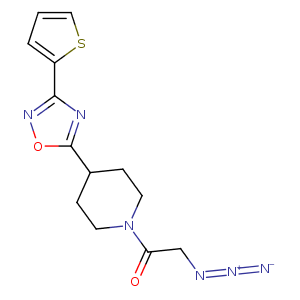
| HET Code: | O8G |
|---|---|
| Formula: | C13H14N6O2S |
| Molecular weight: | 318.354 g/mol |
| DrugBank ID: | - |
| Buried Surface Area: | 71.93 % |
| Polar Surface area: | 137.22 Å2 |
| Number of | |
|---|---|
| H-Bond Acceptors: | 5 |
| H-Bond Donors: | 0 |
| Rings: | 3 |
| Aromatic rings: | 2 |
| Anionic atoms: | 1 |
| Cationic atoms: | 1 |
| Rule of Five Violation: | 0 |
| Rotatable Bonds: | 4 |
| X | Y | Z |
|---|---|---|
| 32.7514 | 71.6316 | 11.8924 |
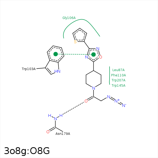


Represent the protein/ligand binding mode, centered on the ligand
Dashed lines represents hydrogen bonds and metal interactions
Green residue labels for amino acids with hydrophobic contacts (green lines) to the ligand
| Ligand | Protein | Interaction | |||
|---|---|---|---|---|---|
| Atom | Atom | Residue | Distance (Å) | Angle (°) | Type |
| C11 | CD2 | LEU- 87 | 3.95 | 0 | Hydrophobic |
| C1 | CG1 | ILE- 107 | 4.07 | 0 | Hydrophobic |
| C11 | CE1 | PHE- 110 | 3.76 | 0 | Hydrophobic |
| S33 | CG2 | VAL- 152 | 4.11 | 0 | Hydrophobic |
| O17 | ND2 | ASN- 179 | 2.97 | 174.97 | H-Bond (Protein Donor) |
| C14 | CH2 | TRP- 207 | 3.98 | 0 | Hydrophobic |
