sc-PDB
An Annotated Database of Druggable Binding Sites from the Protein DataBank
An Annotated Database of Druggable Binding Sites from the Protein DataBank
1.600 Å
X-ray
2009-03-06
| Name: | (S)-mandelate dehydrogenase |
|---|---|
| ID: | MDLB_PSEPU |
| AC: | P20932 |
| Organism: | Pseudomonas putida |
| Reign: | Bacteria |
| TaxID: | 303 |
| EC Number: | 1.1.99.31 |
| Chain Name: | Percentage of Residues within binding site |
|---|---|
| A | 100 % |
| B-Factor: | 13.621 |
|---|---|
| Number of residues: | 49 |
| Including | |
| Standard Amino Acids: | 44 |
| Non Standard Amino Acids: | 0 |
| Water Molecules: | 5 |
| Cofactors: | |
| Metals: | |
| Ligandability | Volume (Å3) |
|---|---|
| 1.031 | 1177.875 |
| % Hydrophobic | % Polar |
|---|---|
| 46.13 | 53.87 |
| According to VolSite | |
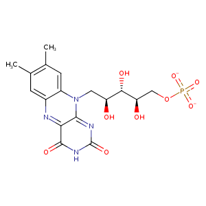
| HET Code: | FMN |
|---|---|
| Formula: | C17H19N4O9P |
| Molecular weight: | 454.328 g/mol |
| DrugBank ID: | DB03247 |
| Buried Surface Area: | 80.55 % |
| Polar Surface area: | 217.05 Å2 |
| Number of | |
|---|---|
| H-Bond Acceptors: | 12 |
| H-Bond Donors: | 4 |
| Rings: | 3 |
| Aromatic rings: | 1 |
| Anionic atoms: | 2 |
| Cationic atoms: | 0 |
| Rule of Five Violation: | 1 |
| Rotatable Bonds: | 7 |
| X | Y | Z |
|---|---|---|
| 75.4316 | 82.7067 | 23.919 |
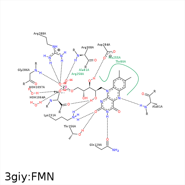


Represent the protein/ligand binding mode, centered on the ligand
Dashed lines represents hydrogen bonds and metal interactions
Green residue labels for amino acids with hydrophobic contacts (green lines) to the ligand
| Ligand | Protein | Interaction | |||
|---|---|---|---|---|---|
| Atom | Atom | Residue | Distance (Å) | Angle (°) | Type |
| C7M | CE2 | TYR- 26 | 3.73 | 0 | Hydrophobic |
| C7M | CD1 | LEU- 27 | 4.04 | 0 | Hydrophobic |
| C8M | CD1 | LEU- 27 | 4.28 | 0 | Hydrophobic |
| O2' | O | PRO- 79 | 2.7 | 168.52 | H-Bond (Ligand Donor) |
| C2' | CG2 | THR- 80 | 4.22 | 0 | Hydrophobic |
| C6 | CB | THR- 80 | 3.8 | 0 | Hydrophobic |
| C9A | CG2 | THR- 80 | 3.77 | 0 | Hydrophobic |
| N5 | N | ALA- 81 | 2.86 | 150.61 | H-Bond (Protein Donor) |
| N3 | OE1 | GLN- 129 | 2.89 | 156.13 | H-Bond (Ligand Donor) |
| O2 | OG1 | THR- 156 | 2.63 | 162.83 | H-Bond (Protein Donor) |
| O2 | NZ | LYS- 231 | 2.93 | 144.41 | H-Bond (Protein Donor) |
| O2' | NZ | LYS- 231 | 2.93 | 158.07 | H-Bond (Protein Donor) |
| C8 | CD | ARG- 258 | 3.65 | 0 | Hydrophobic |
| C9 | CD | ARG- 258 | 3.57 | 0 | Hydrophobic |
| O3' | OD2 | ASP- 284 | 2.83 | 167.35 | H-Bond (Ligand Donor) |
| O3' | OD1 | ASP- 284 | 3.32 | 129.29 | H-Bond (Ligand Donor) |
| C5' | CB | ASP- 284 | 4.02 | 0 | Hydrophobic |
| C5' | CB | SER- 285 | 4.4 | 0 | Hydrophobic |
| O1P | N | GLY- 286 | 2.83 | 155.47 | H-Bond (Protein Donor) |
| O1P | CZ | ARG- 288 | 3.32 | 0 | Ionic (Protein Cationic) |
| O1P | NH1 | ARG- 288 | 2.86 | 147.01 | H-Bond (Protein Donor) |
| O1P | NH2 | ARG- 288 | 2.93 | 143.32 | H-Bond (Protein Donor) |
| O3P | N | GLY- 307 | 3.24 | 122.46 | H-Bond (Protein Donor) |
| C8M | CG | ARG- 308 | 3.46 | 0 | Hydrophobic |
| O2P | N | ARG- 308 | 3.11 | 160.68 | H-Bond (Protein Donor) |
| O2P | CZ | ARG- 308 | 3.86 | 0 | Ionic (Protein Cationic) |
| C8M | CD1 | LEU- 311 | 4.48 | 0 | Hydrophobic |
| C7M | CD1 | LEU- 311 | 4.16 | 0 | Hydrophobic |
| O3P | O | HOH- 1064 | 2.83 | 139.53 | H-Bond (Protein Donor) |
| O3P | O | HOH- 1097 | 2.7 | 155.32 | H-Bond (Protein Donor) |
