sc-PDB
An Annotated Database of Druggable Binding Sites from the Protein DataBank
An Annotated Database of Druggable Binding Sites from the Protein DataBank
2.250 Å
X-ray
2008-06-16
| Name: | Thioredoxin reductase 2, mitochondrial |
|---|---|
| ID: | TRXR2_MOUSE |
| AC: | Q9JLT4 |
| Organism: | Mus musculus |
| Reign: | Eukaryota |
| TaxID: | 10090 |
| EC Number: | 1.8.1.9 |
| Chain Name: | Percentage of Residues within binding site |
|---|---|
| A | 100 % |
| B-Factor: | 37.925 |
|---|---|
| Number of residues: | 73 |
| Including | |
| Standard Amino Acids: | 65 |
| Non Standard Amino Acids: | 0 |
| Water Molecules: | 8 |
| Cofactors: | |
| Metals: | |
| Ligandability | Volume (Å3) |
|---|---|
| 1.081 | 1542.375 |
| % Hydrophobic | % Polar |
|---|---|
| 42.45 | 57.55 |
| According to VolSite | |
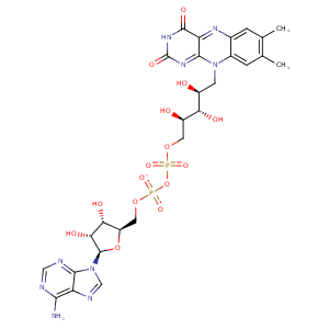
| HET Code: | FAD |
|---|---|
| Formula: | C27H31N9O15P2 |
| Molecular weight: | 783.534 g/mol |
| DrugBank ID: | DB03147 |
| Buried Surface Area: | 73.52 % |
| Polar Surface area: | 381.7 Å2 |
| Number of | |
|---|---|
| H-Bond Acceptors: | 22 |
| H-Bond Donors: | 7 |
| Rings: | 6 |
| Aromatic rings: | 3 |
| Anionic atoms: | 2 |
| Cationic atoms: | 0 |
| Rule of Five Violation: | 3 |
| Rotatable Bonds: | 13 |
| X | Y | Z |
|---|---|---|
| 91.5559 | 102.43 | -50.123 |


Represent the protein/ligand binding mode, centered on the ligand
Dashed lines represents hydrogen bonds and metal interactions
Green residue labels for amino acids with hydrophobic contacts (green lines) to the ligand
| Ligand | Protein | Interaction | |||
|---|---|---|---|---|---|
| Atom | Atom | Residue | Distance (Å) | Angle (°) | Type |
| C4' | CB | SER- 16 | 4.28 | 0 | Hydrophobic |
| O1P | N | GLY- 17 | 2.72 | 160.69 | H-Bond (Protein Donor) |
| O3B | OD1 | ASP- 36 | 2.66 | 157.25 | H-Bond (Ligand Donor) |
| O2B | O | TYR- 37 | 3.09 | 169.54 | H-Bond (Ligand Donor) |
| N3A | N | TYR- 37 | 3.06 | 140.93 | H-Bond (Protein Donor) |
| O1A | N | THR- 52 | 2.78 | 157.32 | H-Bond (Protein Donor) |
| C8M | CG2 | THR- 52 | 3.95 | 0 | Hydrophobic |
| C2' | CB | CYS- 53 | 4.44 | 0 | Hydrophobic |
| O4' | N | CYS- 53 | 3.42 | 138.25 | H-Bond (Protein Donor) |
| C9A | SG | CYS- 58 | 4.41 | 0 | Hydrophobic |
| C2' | SG | CYS- 58 | 4.16 | 0 | Hydrophobic |
| O4 | NZ | LYS- 61 | 2.65 | 137.41 | H-Bond (Protein Donor) |
| N5 | NZ | LYS- 61 | 2.82 | 128.07 | H-Bond (Protein Donor) |
| C6 | CG | LYS- 61 | 4.46 | 0 | Hydrophobic |
| N6A | O | ALA- 126 | 2.87 | 157.18 | H-Bond (Ligand Donor) |
| N1A | N | ALA- 126 | 3.11 | 152.98 | H-Bond (Protein Donor) |
| C7M | CB | SER- 175 | 3.89 | 0 | Hydrophobic |
| C7M | CE2 | PHE- 179 | 4.08 | 0 | Hydrophobic |
| C7M | CG1 | VAL- 196 | 4.04 | 0 | Hydrophobic |
| C7 | CG2 | VAL- 196 | 4.05 | 0 | Hydrophobic |
| C8M | CD | ARG- 285 | 4.01 | 0 | Hydrophobic |
| O3' | OD1 | ASP- 326 | 2.92 | 174 | H-Bond (Ligand Donor) |
| C5' | CB | ASP- 326 | 4.29 | 0 | Hydrophobic |
| O2P | N | ASP- 326 | 2.99 | 129.7 | H-Bond (Protein Donor) |
| O2 | N | THR- 335 | 3.04 | 163.51 | H-Bond (Protein Donor) |
| C2' | CB | THR- 335 | 4.47 | 0 | Hydrophobic |
| C4' | CB | THR- 335 | 4.43 | 0 | Hydrophobic |
| O1P | O | HOH- 511 | 2.85 | 161.52 | H-Bond (Protein Donor) |
| O2P | O | HOH- 515 | 2.59 | 179.97 | H-Bond (Protein Donor) |
| O3B | O | HOH- 552 | 3.14 | 160.05 | H-Bond (Protein Donor) |
| O2A | O | HOH- 611 | 2.59 | 146.01 | H-Bond (Protein Donor) |
