sc-PDB
An Annotated Database of Druggable Binding Sites from the Protein DataBank
An Annotated Database of Druggable Binding Sites from the Protein DataBank
1.750 Å
X-ray
2008-06-13
| Name: | Thioredoxin reductase 1, mitochondrial |
|---|---|
| ID: | TRXR1_DROME |
| AC: | P91938 |
| Organism: | Drosophila melanogaster |
| Reign: | Eukaryota |
| TaxID: | 7227 |
| EC Number: | 1.8.1.9 |
| Chain Name: | Percentage of Residues within binding site |
|---|---|
| A | 6 % |
| B | 94 % |
| B-Factor: | 7.178 |
|---|---|
| Number of residues: | 76 |
| Including | |
| Standard Amino Acids: | 69 |
| Non Standard Amino Acids: | 0 |
| Water Molecules: | 7 |
| Cofactors: | |
| Metals: | |
| Ligandability | Volume (Å3) |
|---|---|
| 1.024 | 695.250 |
| % Hydrophobic | % Polar |
|---|---|
| 44.17 | 55.83 |
| According to VolSite | |
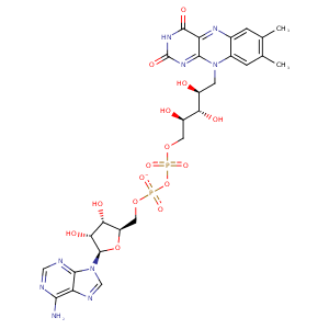
| HET Code: | FAD |
|---|---|
| Formula: | C27H31N9O15P2 |
| Molecular weight: | 783.534 g/mol |
| DrugBank ID: | DB03147 |
| Buried Surface Area: | 75.74 % |
| Polar Surface area: | 381.7 Å2 |
| Number of | |
|---|---|
| H-Bond Acceptors: | 22 |
| H-Bond Donors: | 7 |
| Rings: | 6 |
| Aromatic rings: | 3 |
| Anionic atoms: | 2 |
| Cationic atoms: | 0 |
| Rule of Five Violation: | 3 |
| Rotatable Bonds: | 13 |
| X | Y | Z |
|---|---|---|
| 35.1565 | -13.6605 | 14.0583 |
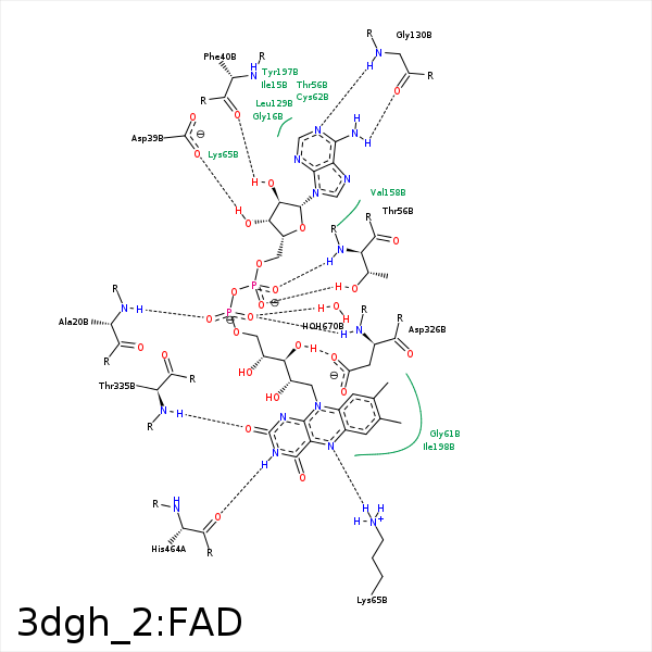


Represent the protein/ligand binding mode, centered on the ligand
Dashed lines represents hydrogen bonds and metal interactions
Green residue labels for amino acids with hydrophobic contacts (green lines) to the ligand
| Ligand | Protein | Interaction | |||
|---|---|---|---|---|---|
| Atom | Atom | Residue | Distance (Å) | Angle (°) | Type |
| C4' | CB | SER- 19 | 4.31 | 0 | Hydrophobic |
| O1P | N | ALA- 20 | 2.88 | 161.91 | H-Bond (Protein Donor) |
| O3B | OD1 | ASP- 39 | 2.84 | 140.73 | H-Bond (Ligand Donor) |
| O3B | OD2 | ASP- 39 | 3.48 | 125.99 | H-Bond (Ligand Donor) |
| O2B | O | PHE- 40 | 2.61 | 152.72 | H-Bond (Ligand Donor) |
| N3A | N | PHE- 40 | 3.24 | 136.91 | H-Bond (Protein Donor) |
| O1A | OG1 | THR- 56 | 2.6 | 165.35 | H-Bond (Protein Donor) |
| O2A | N | THR- 56 | 2.92 | 172.12 | H-Bond (Protein Donor) |
| C8M | CG2 | THR- 56 | 3.48 | 0 | Hydrophobic |
| C2' | CB | CYS- 57 | 4.35 | 0 | Hydrophobic |
| C9A | SG | CYS- 62 | 4.37 | 0 | Hydrophobic |
| C2' | SG | CYS- 62 | 4.28 | 0 | Hydrophobic |
| N5 | NZ | LYS- 65 | 2.98 | 159.47 | H-Bond (Protein Donor) |
| N6A | O | GLY- 130 | 2.97 | 161.4 | H-Bond (Ligand Donor) |
| N1A | N | GLY- 130 | 2.94 | 169.35 | H-Bond (Protein Donor) |
| C7M | CB | SER- 177 | 3.86 | 0 | Hydrophobic |
| C7M | CE2 | PHE- 181 | 4.07 | 0 | Hydrophobic |
| C7M | CG2 | ILE- 198 | 3.81 | 0 | Hydrophobic |
| C7 | CG1 | ILE- 198 | 3.82 | 0 | Hydrophobic |
| C8 | CD1 | ILE- 198 | 3.75 | 0 | Hydrophobic |
| C8M | CD | ARG- 287 | 3.87 | 0 | Hydrophobic |
| O3' | OD2 | ASP- 326 | 2.85 | 172.48 | H-Bond (Ligand Donor) |
| C5' | CB | ASP- 326 | 4.27 | 0 | Hydrophobic |
| O2P | N | ASP- 326 | 2.92 | 155.48 | H-Bond (Protein Donor) |
| N1 | N | THR- 335 | 3.5 | 152.71 | H-Bond (Protein Donor) |
| O2 | N | THR- 335 | 2.94 | 150.9 | H-Bond (Protein Donor) |
| C2' | CB | THR- 335 | 4.4 | 0 | Hydrophobic |
| C4' | CB | THR- 335 | 4.49 | 0 | Hydrophobic |
| C5' | CB | ALA- 338 | 4.33 | 0 | Hydrophobic |
| N3 | O | HIS- 464 | 2.79 | 154.71 | H-Bond (Ligand Donor) |
| O2P | O | HOH- 670 | 2.67 | 170.19 | H-Bond (Protein Donor) |
