sc-PDB
An Annotated Database of Druggable Binding Sites from the Protein DataBank
An Annotated Database of Druggable Binding Sites from the Protein DataBank
1.750 Å
X-ray
2008-05-29
| Name: | 5,10-methenyltetrahydromethanopterin hydrogenase |
|---|---|
| ID: | HMD_METJA |
| AC: | Q58194 |
| Organism: | Methanocaldococcus jannaschii |
| Reign: | Archaea |
| TaxID: | 243232 |
| EC Number: | 1.12.98.2 |
| Chain Name: | Percentage of Residues within binding site |
|---|---|
| A | 100 % |
| B-Factor: | 31.176 |
|---|---|
| Number of residues: | 40 |
| Including | |
| Standard Amino Acids: | 37 |
| Non Standard Amino Acids: | 1 |
| Water Molecules: | 2 |
| Cofactors: | |
| Metals: | FE2 |
| Ligandability | Volume (Å3) |
|---|---|
| 1.084 | 502.875 |
| % Hydrophobic | % Polar |
|---|---|
| 55.70 | 44.30 |
| According to VolSite | |
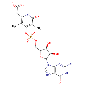
| HET Code: | FEG |
|---|---|
| Formula: | C19H21N6O11P |
| Molecular weight: | 540.377 g/mol |
| DrugBank ID: | - |
| Buried Surface Area: | 64.13 % |
| Polar Surface area: | 276.64 Å2 |
| Number of | |
|---|---|
| H-Bond Acceptors: | 15 |
| H-Bond Donors: | 5 |
| Rings: | 4 |
| Aromatic rings: | 2 |
| Anionic atoms: | 2 |
| Cationic atoms: | 0 |
| Rule of Five Violation: | 2 |
| Rotatable Bonds: | 8 |
| X | Y | Z |
|---|---|---|
| 23.1701 | 9.40665 | 31.7215 |
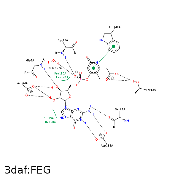


Represent the protein/ligand binding mode, centered on the ligand
Dashed lines represents hydrogen bonds and metal interactions
Green residue labels for amino acids with hydrophobic contacts (green lines) to the ligand
| Ligand | Protein | Interaction | |||
|---|---|---|---|---|---|
| Atom | Atom | Residue | Distance (Å) | Angle (°) | Type |
| O3S | N | GLY- 9 | 2.83 | 134.07 | H-Bond (Protein Donor) |
| O2P | N | CYS- 10 | 3.13 | 157.06 | H-Bond (Protein Donor) |
| O18 | OG1 | THR- 13 | 3.09 | 159.34 | H-Bond (Protein Donor) |
| N2A | O | SER- 63 | 3 | 128.71 | H-Bond (Ligand Donor) |
| O3S | OD2 | ASP- 64 | 2.61 | 145.43 | H-Bond (Ligand Donor) |
| O2S | OD2 | ASP- 64 | 3.16 | 156.27 | H-Bond (Ligand Donor) |
| O2S | OD1 | ASP- 64 | 2.67 | 134.98 | H-Bond (Ligand Donor) |
| C2S | CB | PRO- 114 | 3.69 | 0 | Hydrophobic |
| C2S | CB | PRO- 115 | 4.26 | 0 | Hydrophobic |
| C3S | SG | CYS- 118 | 4.13 | 0 | Hydrophobic |
| N1A | OD2 | ASP- 135 | 2.91 | 169.56 | H-Bond (Ligand Donor) |
| N2A | OD1 | ASP- 135 | 2.83 | 145.25 | H-Bond (Ligand Donor) |
| N2A | OD2 | ASP- 135 | 3.48 | 134.87 | H-Bond (Ligand Donor) |
| C3M | CE2 | TRP- 148 | 3.57 | 0 | Hydrophobic |
| C1S | CG | PRO- 150 | 3.95 | 0 | Hydrophobic |
| C4S | CG | PRO- 150 | 4.32 | 0 | Hydrophobic |
| C7 | SG | CYS- 176 | 4.39 | 0 | Hydrophobic |
| C3M | CG2 | THR- 177 | 3.84 | 0 | Hydrophobic |
| O2P | O | HOH- 2067 | 2.55 | 179.98 | H-Bond (Protein Donor) |
