sc-PDB
An Annotated Database of Druggable Binding Sites from the Protein DataBank
An Annotated Database of Druggable Binding Sites from the Protein DataBank
1.400 Å
X-ray
2008-05-22
| Name: | Dihydrofolate reductase |
|---|---|
| ID: | DYR_MOUSE |
| AC: | P00375 |
| Organism: | Mus musculus |
| Reign: | Eukaryota |
| TaxID: | 10090 |
| EC Number: | 1.5.1.3 |
| Chain Name: | Percentage of Residues within binding site |
|---|---|
| A | 100 % |
| B-Factor: | 13.172 |
|---|---|
| Number of residues: | 28 |
| Including | |
| Standard Amino Acids: | 26 |
| Non Standard Amino Acids: | 1 |
| Water Molecules: | 1 |
| Cofactors: | NDP |
| Metals: | |
| Ligandability | Volume (Å3) |
|---|---|
| 1.034 | 604.125 |
| % Hydrophobic | % Polar |
|---|---|
| 61.45 | 38.55 |
| According to VolSite | |
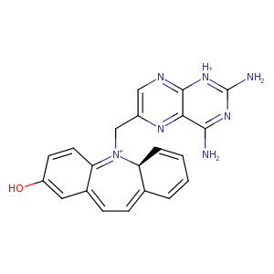
| HET Code: | Q22 |
|---|---|
| Formula: | C21H19N7O |
| Molecular weight: | 385.422 g/mol |
| DrugBank ID: | DB08448 |
| Buried Surface Area: | 70.03 % |
| Polar Surface area: | 127.07 Å2 |
| Number of | |
|---|---|
| H-Bond Acceptors: | 8 |
| H-Bond Donors: | 3 |
| Rings: | 5 |
| Aromatic rings: | 3 |
| Anionic atoms: | 0 |
| Cationic atoms: | 0 |
| Rule of Five Violation: | 0 |
| Rotatable Bonds: | 2 |
| X | Y | Z |
|---|---|---|
| -5.07683 | 2.19697 | 14.8553 |
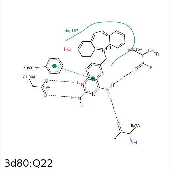


Represent the protein/ligand binding mode, centered on the ligand
Dashed lines represents hydrogen bonds and metal interactions
Green residue labels for amino acids with hydrophobic contacts (green lines) to the ligand
| Ligand | Protein | Interaction | |||
|---|---|---|---|---|---|
| Atom | Atom | Residue | Distance (Å) | Angle (°) | Type |
| N4' | O | ILE- 7 | 2.9 | 156.23 | H-Bond (Ligand Donor) |
| N1' | OE2 | GLU- 30 | 2.82 | 165.42 | H-Bond (Ligand Donor) |
| N2' | OE1 | GLU- 30 | 2.79 | 173.28 | H-Bond (Ligand Donor) |
| N4' | O | VAL- 115 | 3.12 | 134.36 | H-Bond (Ligand Donor) |
