sc-PDB
An Annotated Database of Druggable Binding Sites from the Protein DataBank
An Annotated Database of Druggable Binding Sites from the Protein DataBank
2.540 Å
X-ray
2010-10-26
| Name: | Putative flavin-containing monooxygenase |
|---|---|
| ID: | Q83XK4_9GAMM |
| AC: | Q83XK4 |
| Organism: | Methylophaga aminisulfidivorans |
| Reign: | Bacteria |
| TaxID: | 230105 |
| EC Number: | / |
| Chain Name: | Percentage of Residues within binding site |
|---|---|
| A | 100 % |
| B-Factor: | 36.582 |
|---|---|
| Number of residues: | 60 |
| Including | |
| Standard Amino Acids: | 56 |
| Non Standard Amino Acids: | 1 |
| Water Molecules: | 3 |
| Cofactors: | NAP |
| Metals: | |
| Ligandability | Volume (Å3) |
|---|---|
| 0.897 | 847.125 |
| % Hydrophobic | % Polar |
|---|---|
| 37.85 | 62.15 |
| According to VolSite | |
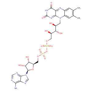
| HET Code: | FAD |
|---|---|
| Formula: | C27H31N9O15P2 |
| Molecular weight: | 783.534 g/mol |
| DrugBank ID: | DB03147 |
| Buried Surface Area: | 76.38 % |
| Polar Surface area: | 381.7 Å2 |
| Number of | |
|---|---|
| H-Bond Acceptors: | 22 |
| H-Bond Donors: | 7 |
| Rings: | 6 |
| Aromatic rings: | 3 |
| Anionic atoms: | 2 |
| Cationic atoms: | 0 |
| Rule of Five Violation: | 3 |
| Rotatable Bonds: | 13 |
| X | Y | Z |
|---|---|---|
| 31.0637 | 26.6695 | -43.8739 |


Represent the protein/ligand binding mode, centered on the ligand
Dashed lines represents hydrogen bonds and metal interactions
Green residue labels for amino acids with hydrophobic contacts (green lines) to the ligand
| Ligand | Protein | Interaction | |||
|---|---|---|---|---|---|
| Atom | Atom | Residue | Distance (Å) | Angle (°) | Type |
| C4' | CG | PRO- 12 | 3.67 | 0 | Hydrophobic |
| O1P | N | SER- 13 | 2.85 | 159.95 | H-Bond (Protein Donor) |
| O2P | OG | SER- 13 | 2.86 | 157.19 | H-Bond (Protein Donor) |
| O3B | OE1 | GLU- 38 | 2.78 | 167.79 | H-Bond (Ligand Donor) |
| N3A | N | LYS- 39 | 3.44 | 149.61 | H-Bond (Protein Donor) |
| O2B | NE2 | GLN- 40 | 3.06 | 172.91 | H-Bond (Protein Donor) |
| O2A | N | GLN- 46 | 2.82 | 179.05 | H-Bond (Protein Donor) |
| C8M | CG | GLN- 46 | 4.13 | 0 | Hydrophobic |
| O3' | NE1 | TRP- 47 | 2.98 | 147.47 | H-Bond (Protein Donor) |
| O4' | NE1 | TRP- 47 | 3.36 | 121.09 | H-Bond (Protein Donor) |
| O1A | NE2 | HIS- 63 | 2.57 | 152 | H-Bond (Protein Donor) |
| O5B | NE2 | HIS- 63 | 3.42 | 125.24 | H-Bond (Protein Donor) |
| C2B | CB | HIS- 63 | 4.06 | 0 | Hydrophobic |
| C7M | CB | SER- 65 | 4.15 | 0 | Hydrophobic |
| C8M | CB | SER- 65 | 3.85 | 0 | Hydrophobic |
| C7M | CG | MET- 66 | 3.93 | 0 | Hydrophobic |
| C6 | CE | MET- 66 | 3.55 | 0 | Hydrophobic |
| C7M | CD1 | LEU- 70 | 3.89 | 0 | Hydrophobic |
| O4 | N | ASN- 73 | 2.95 | 156.38 | H-Bond (Protein Donor) |
| N6A | O | VAL- 126 | 3.43 | 164.66 | H-Bond (Ligand Donor) |
| N1A | N | VAL- 126 | 2.95 | 175.21 | H-Bond (Protein Donor) |
| C8M | CB | PHE- 165 | 3.61 | 0 | Hydrophobic |
| C2' | CD2 | PHE- 165 | 4.42 | 0 | Hydrophobic |
| O2' | OE1 | GLN- 318 | 2.86 | 159.28 | H-Bond (Ligand Donor) |
| O2 | OG | SER- 321 | 2.57 | 146.61 | H-Bond (Protein Donor) |
| C5' | CE2 | PHE- 325 | 3.62 | 0 | Hydrophobic |
| N5 | N7N | NAP- 1449 | 2.9 | 162.23 | H-Bond (Protein Donor) |
| C7M | C5N | NAP- 1449 | 3.97 | 0 | Hydrophobic |
| C9 | C4D | NAP- 1449 | 4.44 | 0 | Hydrophobic |
| O2P | O | HOH- 2038 | 2.7 | 155.86 | H-Bond (Protein Donor) |
| O1A | O | HOH- 2054 | 2.67 | 179.96 | H-Bond (Protein Donor) |
