sc-PDB
An Annotated Database of Druggable Binding Sites from the Protein DataBank
An Annotated Database of Druggable Binding Sites from the Protein DataBank
1.900 Å
X-ray
2009-03-20
| Name: | Pteridine reductase |
|---|---|
| ID: | O76290_TRYBB |
| AC: | O76290 |
| Organism: | Trypanosoma brucei brucei |
| Reign: | Eukaryota |
| TaxID: | 5702 |
| EC Number: | / |
| Chain Name: | Percentage of Residues within binding site |
|---|---|
| C | 100 % |
| B-Factor: | 27.069 |
|---|---|
| Number of residues: | 20 |
| Including | |
| Standard Amino Acids: | 19 |
| Non Standard Amino Acids: | 1 |
| Water Molecules: | 0 |
| Cofactors: | NAP |
| Metals: | |
| Ligandability | Volume (Å3) |
|---|---|
| 0.786 | 793.125 |
| % Hydrophobic | % Polar |
|---|---|
| 50.21 | 49.79 |
| According to VolSite | |
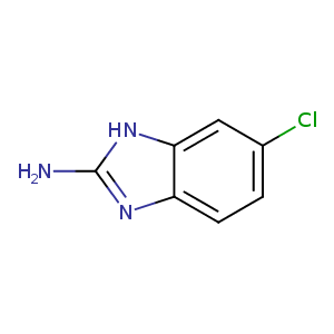
| HET Code: | VGD |
|---|---|
| Formula: | C7H6ClN3 |
| Molecular weight: | 167.596 g/mol |
| DrugBank ID: | - |
| Buried Surface Area: | 72.12 % |
| Polar Surface area: | 54.7 Å2 |
| Number of | |
|---|---|
| H-Bond Acceptors: | 2 |
| H-Bond Donors: | 2 |
| Rings: | 2 |
| Aromatic rings: | 2 |
| Anionic atoms: | 0 |
| Cationic atoms: | 0 |
| Rule of Five Violation: | 0 |
| Rotatable Bonds: | 0 |
| X | Y | Z |
|---|---|---|
| 1.852 | 16.4827 | 32.0673 |
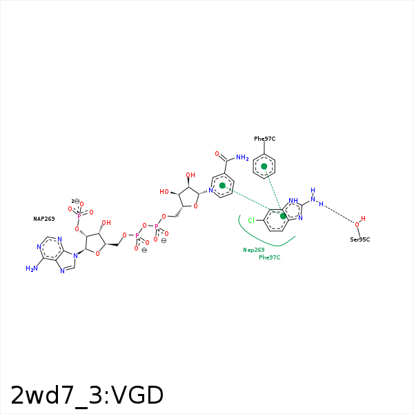


Represent the protein/ligand binding mode, centered on the ligand
Dashed lines represents hydrogen bonds and metal interactions
Green residue labels for amino acids with hydrophobic contacts (green lines) to the ligand
| Ligand | Protein | Interaction | |||
|---|---|---|---|---|---|
| Atom | Atom | Residue | Distance (Å) | Angle (°) | Type |
| NAA | OG | SER- 95 | 2.71 | 164.9 | H-Bond (Ligand Donor) |
| CAJ | CB | PHE- 97 | 4.17 | 0 | Hydrophobic |
| DuAr | DuAr | PHE- 97 | 3.63 | 0 | Aromatic Face/Face |
| CLA | CD2 | LEU- 209 | 4.15 | 0 | Hydrophobic |
| CAE | CG | PRO- 210 | 4.02 | 0 | Hydrophobic |
| CLA | CG | PRO- 210 | 4.16 | 0 | Hydrophobic |
| CLA | C4N | NAP- 269 | 4.35 | 0 | Hydrophobic |
| CAJ | C2D | NAP- 269 | 4.46 | 0 | Hydrophobic |
| NAF | O2A | NAP- 269 | 3.31 | 137.93 | H-Bond (Ligand Donor) |
| NAA | O2A | NAP- 269 | 3.3 | 149.02 | H-Bond (Ligand Donor) |
