sc-PDB
An Annotated Database of Druggable Binding Sites from the Protein DataBank
An Annotated Database of Druggable Binding Sites from the Protein DataBank
1.930 Å
X-ray
2008-02-05
| Name: | NADPH:ferredoxin reductase |
|---|---|
| ID: | Q9L6V3_RHOCA |
| AC: | Q9L6V3 |
| Organism: | Rhodobacter capsulatus |
| Reign: | Bacteria |
| TaxID: | 1061 |
| EC Number: | / |
| Chain Name: | Percentage of Residues within binding site |
|---|---|
| C | 100 % |
| B-Factor: | 16.473 |
|---|---|
| Number of residues: | 41 |
| Including | |
| Standard Amino Acids: | 37 |
| Non Standard Amino Acids: | 1 |
| Water Molecules: | 3 |
| Cofactors: | NAP |
| Metals: | |
| Ligandability | Volume (Å3) |
|---|---|
| 0.909 | 756.000 |
| % Hydrophobic | % Polar |
|---|---|
| 45.54 | 54.46 |
| According to VolSite | |
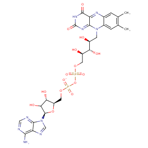
| HET Code: | FAD |
|---|---|
| Formula: | C27H31N9O15P2 |
| Molecular weight: | 783.534 g/mol |
| DrugBank ID: | DB03147 |
| Buried Surface Area: | 66.18 % |
| Polar Surface area: | 381.7 Å2 |
| Number of | |
|---|---|
| H-Bond Acceptors: | 22 |
| H-Bond Donors: | 7 |
| Rings: | 6 |
| Aromatic rings: | 3 |
| Anionic atoms: | 2 |
| Cationic atoms: | 0 |
| Rule of Five Violation: | 3 |
| Rotatable Bonds: | 13 |
| X | Y | Z |
|---|---|---|
| 32.0019 | 28.2951 | -4.34898 |
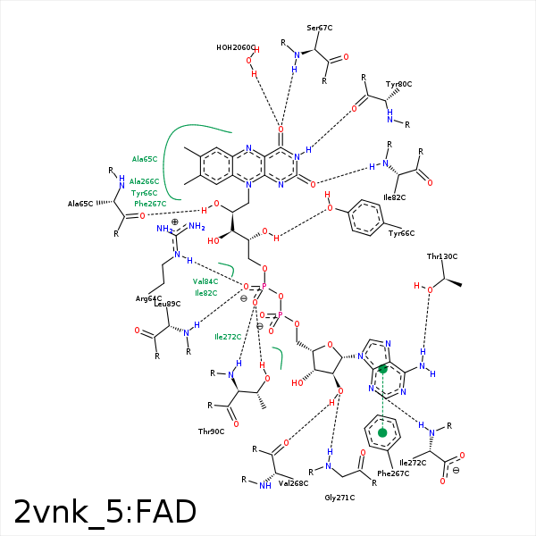


Represent the protein/ligand binding mode, centered on the ligand
Dashed lines represents hydrogen bonds and metal interactions
Green residue labels for amino acids with hydrophobic contacts (green lines) to the ligand
| Ligand | Protein | Interaction | |||
|---|---|---|---|---|---|
| Atom | Atom | Residue | Distance (Å) | Angle (°) | Type |
| C7M | CE1 | PHE- 49 | 3.93 | 0 | Hydrophobic |
| C2' | CB | ARG- 64 | 4.07 | 0 | Hydrophobic |
| C3' | CD | ARG- 64 | 3.93 | 0 | Hydrophobic |
| O1P | NH2 | ARG- 64 | 3.23 | 132.27 | H-Bond (Protein Donor) |
| O1P | NE | ARG- 64 | 2.89 | 148.64 | H-Bond (Protein Donor) |
| O1P | CZ | ARG- 64 | 3.47 | 0 | Ionic (Protein Cationic) |
| O2' | O | ALA- 65 | 2.79 | 166.83 | H-Bond (Ligand Donor) |
| C7 | CB | ALA- 65 | 3.66 | 0 | Hydrophobic |
| C8 | CB | ALA- 65 | 3.71 | 0 | Hydrophobic |
| C8 | CB | ALA- 65 | 3.71 | 0 | Hydrophobic |
| C2' | CE1 | TYR- 66 | 4.28 | 0 | Hydrophobic |
| C4' | CE1 | TYR- 66 | 4.36 | 0 | Hydrophobic |
| O4' | OH | TYR- 66 | 2.78 | 135.09 | H-Bond (Ligand Donor) |
| O4 | N | SER- 67 | 3.16 | 140.3 | H-Bond (Protein Donor) |
| N3 | O | TYR- 80 | 2.85 | 168.94 | H-Bond (Ligand Donor) |
| O2 | N | ILE- 82 | 2.89 | 167.12 | H-Bond (Protein Donor) |
| C5' | CG2 | ILE- 82 | 4.06 | 0 | Hydrophobic |
| C5' | CG2 | VAL- 84 | 4.37 | 0 | Hydrophobic |
| C5B | CG1 | VAL- 84 | 4.1 | 0 | Hydrophobic |
| O1P | N | LEU- 89 | 3.03 | 170.5 | H-Bond (Protein Donor) |
| O2P | N | LEU- 89 | 3.47 | 121.07 | H-Bond (Protein Donor) |
| O2P | OG1 | THR- 90 | 2.7 | 176.41 | H-Bond (Protein Donor) |
| O2P | N | THR- 90 | 2.86 | 164.31 | H-Bond (Protein Donor) |
| C5' | CG2 | THR- 90 | 3.77 | 0 | Hydrophobic |
| N6A | OG1 | THR- 130 | 2.83 | 159.17 | H-Bond (Ligand Donor) |
| C7M | CG | GLU- 264 | 4.08 | 0 | Hydrophobic |
| C1' | CB | PHE- 267 | 3.84 | 0 | Hydrophobic |
| C2B | CD1 | PHE- 267 | 4.05 | 0 | Hydrophobic |
| DuAr | DuAr | PHE- 267 | 3.79 | 0 | Aromatic Face/Face |
| O2B | O | VAL- 268 | 3.08 | 157.67 | H-Bond (Ligand Donor) |
| C8M | CG2 | VAL- 268 | 4.19 | 0 | Hydrophobic |
| O2B | N | GLY- 271 | 3.02 | 135.11 | H-Bond (Protein Donor) |
| C4B | CD1 | ILE- 272 | 3.99 | 0 | Hydrophobic |
| C1B | CG1 | ILE- 272 | 3.72 | 0 | Hydrophobic |
| N3A | N | ILE- 272 | 3.19 | 167.52 | H-Bond (Protein Donor) |
| O4 | O | HOH- 2060 | 2.92 | 156.24 | H-Bond (Protein Donor) |
