sc-PDB
An Annotated Database of Druggable Binding Sites from the Protein DataBank
An Annotated Database of Druggable Binding Sites from the Protein DataBank
2.000 Å
X-ray
2007-11-26
| Name: | Alanine dehydrogenase |
|---|---|
| ID: | DHA_MYCTU |
| AC: | P9WQB1 |
| Organism: | Mycobacterium tuberculosis |
| Reign: | Bacteria |
| TaxID: | 83332 |
| EC Number: | 1.4.1.1 |
| Chain Name: | Percentage of Residues within binding site |
|---|---|
| E | 100 % |
| B-Factor: | 43.370 |
|---|---|
| Number of residues: | 53 |
| Including | |
| Standard Amino Acids: | 52 |
| Non Standard Amino Acids: | 0 |
| Water Molecules: | 1 |
| Cofactors: | |
| Metals: | |
| Ligandability | Volume (Å3) |
|---|---|
| 0.567 | 472.500 |
| % Hydrophobic | % Polar |
|---|---|
| 48.57 | 51.43 |
| According to VolSite | |
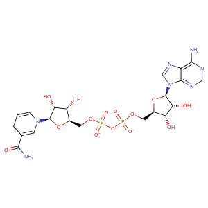
| HET Code: | NAI |
|---|---|
| Formula: | C21H27N7O14P2 |
| Molecular weight: | 663.425 g/mol |
| DrugBank ID: | DB00157 |
| Buried Surface Area: | 70.19 % |
| Polar Surface area: | 342.9 Å2 |
| Number of | |
|---|---|
| H-Bond Acceptors: | 19 |
| H-Bond Donors: | 6 |
| Rings: | 5 |
| Aromatic rings: | 2 |
| Anionic atoms: | 2 |
| Cationic atoms: | 0 |
| Rule of Five Violation: | 3 |
| Rotatable Bonds: | 11 |
| X | Y | Z |
|---|---|---|
| -74.3054 | 38.3107 | 15.8215 |
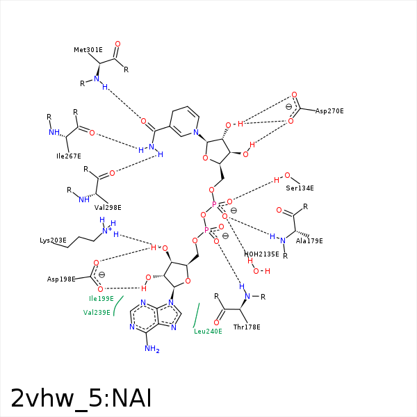


Represent the protein/ligand binding mode, centered on the ligand
Dashed lines represents hydrogen bonds and metal interactions
Green residue labels for amino acids with hydrophobic contacts (green lines) to the ligand
| Ligand | Protein | Interaction | |||
|---|---|---|---|---|---|
| Atom | Atom | Residue | Distance (Å) | Angle (°) | Type |
| C2D | CD2 | LEU- 130 | 3.97 | 0 | Hydrophobic |
| C4N | CB | MET- 133 | 4.44 | 0 | Hydrophobic |
| C4N | CB | ALA- 137 | 3.5 | 0 | Hydrophobic |
| O2A | N | THR- 178 | 3.12 | 162.44 | H-Bond (Protein Donor) |
| O2N | N | ALA- 179 | 2.86 | 160.82 | H-Bond (Protein Donor) |
| C5D | CB | ALA- 179 | 4.18 | 0 | Hydrophobic |
| O3B | OD2 | ASP- 198 | 2.7 | 138.07 | H-Bond (Ligand Donor) |
| O2B | OD1 | ASP- 198 | 2.67 | 163.76 | H-Bond (Ligand Donor) |
| O2B | OD2 | ASP- 198 | 3.41 | 130.24 | H-Bond (Ligand Donor) |
| C3B | CD | LYS- 203 | 4.41 | 0 | Hydrophobic |
| O3B | NZ | LYS- 203 | 2.95 | 163.66 | H-Bond (Protein Donor) |
| C1B | CB | VAL- 239 | 4.47 | 0 | Hydrophobic |
| C5B | CB | LEU- 240 | 3.87 | 0 | Hydrophobic |
| C3D | CD1 | LEU- 240 | 3.73 | 0 | Hydrophobic |
| C5D | CB | LEU- 240 | 3.87 | 0 | Hydrophobic |
| O4B | N | LEU- 240 | 3.38 | 150.64 | H-Bond (Protein Donor) |
| C1D | CG2 | ILE- 267 | 3.96 | 0 | Hydrophobic |
| N7N | O | ILE- 267 | 3.12 | 174.48 | H-Bond (Ligand Donor) |
| O3D | OD2 | ASP- 270 | 2.96 | 158.71 | H-Bond (Ligand Donor) |
| O2D | OD2 | ASP- 270 | 3.03 | 151.2 | H-Bond (Ligand Donor) |
| N7N | O | VAL- 298 | 3.05 | 165.61 | H-Bond (Ligand Donor) |
| O7N | N | MET- 301 | 2.74 | 147.37 | H-Bond (Protein Donor) |
| C4N | CG | PRO- 302 | 4.27 | 0 | Hydrophobic |
| O2N | O | HOH- 2135 | 2.69 | 179.96 | H-Bond (Protein Donor) |
