sc-PDB
An Annotated Database of Druggable Binding Sites from the Protein DataBank
An Annotated Database of Druggable Binding Sites from the Protein DataBank
1.700 Å
X-ray
2007-11-15
| Name: | Aurora kinase B-A |
|---|---|
| ID: | AUKBA_XENLA |
| AC: | Q6DE08 |
| Organism: | Xenopus laevis |
| Reign: | Eukaryota |
| TaxID: | 8355 |
| EC Number: | 2.7.11.1 |
| Chain Name: | Percentage of Residues within binding site |
|---|---|
| B | 100 % |
| B-Factor: | 25.565 |
|---|---|
| Number of residues: | 26 |
| Including | |
| Standard Amino Acids: | 25 |
| Non Standard Amino Acids: | 0 |
| Water Molecules: | 1 |
| Cofactors: | |
| Metals: | |
| Ligandability | Volume (Å3) |
|---|---|
| 0.755 | 526.500 |
| % Hydrophobic | % Polar |
|---|---|
| 44.87 | 55.13 |
| According to VolSite | |
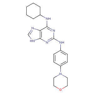
| HET Code: | AD5 |
|---|---|
| Formula: | C21H27N7O |
| Molecular weight: | 393.485 g/mol |
| DrugBank ID: | DB07340 |
| Buried Surface Area: | 45.69 % |
| Polar Surface area: | 90.99 Å2 |
| Number of | |
|---|---|
| H-Bond Acceptors: | 7 |
| H-Bond Donors: | 3 |
| Rings: | 5 |
| Aromatic rings: | 3 |
| Anionic atoms: | 0 |
| Cationic atoms: | 0 |
| Rule of Five Violation: | 0 |
| Rotatable Bonds: | 5 |
| X | Y | Z |
|---|---|---|
| 40.2567 | 51.5116 | 58.0546 |
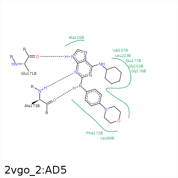


Represent the protein/ligand binding mode, centered on the ligand
Dashed lines represents hydrogen bonds and metal interactions
Green residue labels for amino acids with hydrophobic contacts (green lines) to the ligand
| Ligand | Protein | Interaction | |||
|---|---|---|---|---|---|
| Atom | Atom | Residue | Distance (Å) | Angle (°) | Type |
| CAL | CG2 | VAL- 107 | 3.84 | 0 | Hydrophobic |
| N9 | O | GLU- 171 | 2.89 | 162.09 | H-Bond (Ligand Donor) |
| N3 | N | ALA- 173 | 3.15 | 167.27 | H-Bond (Protein Donor) |
| N2 | O | ALA- 173 | 2.64 | 131.62 | H-Bond (Ligand Donor) |
| CAG | CG | GLU- 177 | 3.74 | 0 | Hydrophobic |
| CAK | CD2 | LEU- 223 | 4.07 | 0 | Hydrophobic |
