sc-PDB
An Annotated Database of Druggable Binding Sites from the Protein DataBank
An Annotated Database of Druggable Binding Sites from the Protein DataBank
2.170 Å
X-ray
2006-09-27
| Name: | Lipoamide acyltransferase component of branched-chain alpha-keto acid dehydrogenase complex, mitochondrial |
|---|---|
| ID: | ODB2_BOVIN |
| AC: | P11181 |
| Organism: | Bos taurus |
| Reign: | Eukaryota |
| TaxID: | 9913 |
| EC Number: | 2.3.1.168 |
| Chain Name: | Percentage of Residues within binding site |
|---|---|
| E | 85 % |
| F | 15 % |
| B-Factor: | 39.004 |
|---|---|
| Number of residues: | 42 |
| Including | |
| Standard Amino Acids: | 41 |
| Non Standard Amino Acids: | 0 |
| Water Molecules: | 1 |
| Cofactors: | |
| Metals: | |
| Ligandability | Volume (Å3) |
|---|---|
| 0.059 | 330.750 |
| % Hydrophobic | % Polar |
|---|---|
| 42.86 | 57.14 |
| According to VolSite | |
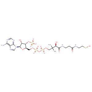
| HET Code: | CAO |
|---|---|
| Formula: | C21H32N7O17P3S |
| Molecular weight: | 779.502 g/mol |
| DrugBank ID: | DB01846 |
| Buried Surface Area: | 56.84 % |
| Polar Surface area: | 432.84 Å2 |
| Number of | |
|---|---|
| H-Bond Acceptors: | 22 |
| H-Bond Donors: | 6 |
| Rings: | 3 |
| Aromatic rings: | 2 |
| Anionic atoms: | 4 |
| Cationic atoms: | 0 |
| Rule of Five Violation: | 3 |
| Rotatable Bonds: | 19 |
| X | Y | Z |
|---|---|---|
| 90.0345 | -12.0055 | 30.9371 |
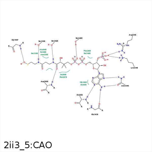


Represent the protein/ligand binding mode, centered on the ligand
Dashed lines represents hydrogen bonds and metal interactions
Green residue labels for amino acids with hydrophobic contacts (green lines) to the ligand
| Ligand | Protein | Interaction | |||
|---|---|---|---|---|---|
| Atom | Atom | Residue | Distance (Å) | Angle (°) | Type |
| S1P | CE1 | PHE- 215 | 3.93 | 0 | Hydrophobic |
| O8A | CZ | ARG- 230 | 3.26 | 0 | Ionic (Protein Cationic) |
| O8A | NZ | LYS- 234 | 2.89 | 157.29 | H-Bond (Protein Donor) |
| O8A | NZ | LYS- 234 | 2.89 | 0 | Ionic (Protein Cationic) |
| C5B | CB | SER- 245 | 4.21 | 0 | Hydrophobic |
| O4A | OG | SER- 245 | 3.42 | 132.14 | H-Bond (Protein Donor) |
| O5A | OG | SER- 245 | 2.81 | 158.7 | H-Bond (Protein Donor) |
| C2B | CD2 | PHE- 246 | 4.35 | 0 | Hydrophobic |
| CDP | CG2 | ILE- 285 | 3.35 | 0 | Hydrophobic |
| C2P | CB | ALA- 286 | 3.95 | 0 | Hydrophobic |
| CEP | CB | MET- 287 | 3.7 | 0 | Hydrophobic |
| O9P | N | ASP- 288 | 2.92 | 169.33 | H-Bond (Protein Donor) |
| C2P | CD1 | LEU- 293 | 3.6 | 0 | Hydrophobic |
| S1P | CD2 | LEU- 293 | 3.77 | 0 | Hydrophobic |
| O1A | NE2 | GLN- 317 | 3.35 | 145.14 | H-Bond (Protein Donor) |
| CEP | CG | GLN- 317 | 4.08 | 0 | Hydrophobic |
| N4P | OG | SER- 338 | 2.65 | 133.4 | H-Bond (Ligand Donor) |
| C2P | CB | SER- 338 | 4.23 | 0 | Hydrophobic |
| N6A | OD1 | ASN- 339 | 3.17 | 157.65 | H-Bond (Ligand Donor) |
| N7A | ND2 | ASN- 339 | 2.92 | 170.3 | H-Bond (Protein Donor) |
| CDP | CB | ASN- 339 | 4.05 | 0 | Hydrophobic |
| CAP | CB | SER- 342 | 4.11 | 0 | Hydrophobic |
| N6A | O | GLY- 363 | 2.73 | 153.44 | H-Bond (Ligand Donor) |
| N1A | N | ILE- 365 | 2.87 | 159.9 | H-Bond (Protein Donor) |
| O1P | N | GLY- 396 | 3.12 | 176.34 | H-Bond (Protein Donor) |
