sc-PDB
An Annotated Database of Druggable Binding Sites from the Protein DataBank
An Annotated Database of Druggable Binding Sites from the Protein DataBank
2.500 Å
X-ray
2006-09-15
| Name: | Cob(I)yrinic acid a,c-diamide adenosyltransferase, mitochondrial |
|---|---|
| ID: | MMAB_HUMAN |
| AC: | Q96EY8 |
| Organism: | Homo sapiens |
| Reign: | Eukaryota |
| TaxID: | 9606 |
| EC Number: | 2.5.1.17 |
| Chain Name: | Percentage of Residues within binding site |
|---|---|
| B | 65 % |
| C | 35 % |
| B-Factor: | 24.211 |
|---|---|
| Number of residues: | 35 |
| Including | |
| Standard Amino Acids: | 32 |
| Non Standard Amino Acids: | 2 |
| Water Molecules: | 1 |
| Cofactors: | |
| Metals: | MG MG |
| Ligandability | Volume (Å3) |
|---|---|
| 0.100 | 779.625 |
| % Hydrophobic | % Polar |
|---|---|
| 43.29 | 56.71 |
| According to VolSite | |

| HET Code: | ATP |
|---|---|
| Formula: | C10H12N5O13P3 |
| Molecular weight: | 503.149 g/mol |
| DrugBank ID: | DB00171 |
| Buried Surface Area: | 63.41 % |
| Polar Surface area: | 319.88 Å2 |
| Number of | |
|---|---|
| H-Bond Acceptors: | 17 |
| H-Bond Donors: | 3 |
| Rings: | 3 |
| Aromatic rings: | 2 |
| Anionic atoms: | 4 |
| Cationic atoms: | 0 |
| Rule of Five Violation: | 2 |
| Rotatable Bonds: | 8 |
| X | Y | Z |
|---|---|---|
| 44.7649 | 44.9575 | 8.5759 |
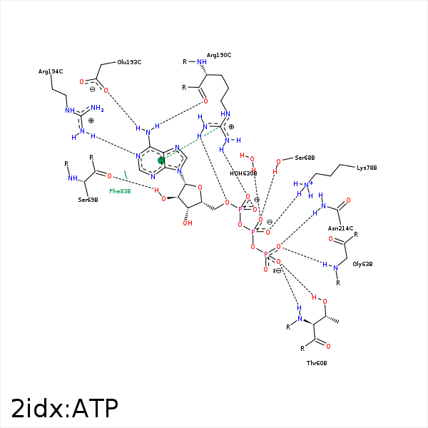


Represent the protein/ligand binding mode, centered on the ligand
Dashed lines represents hydrogen bonds and metal interactions
Green residue labels for amino acids with hydrophobic contacts (green lines) to the ligand
| Ligand | Protein | Interaction | |||
|---|---|---|---|---|---|
| Atom | Atom | Residue | Distance (Å) | Angle (°) | Type |
| O2G | N | THR- 60 | 2.8 | 162.67 | H-Bond (Protein Donor) |
| O2G | OG1 | THR- 60 | 2.61 | 160.37 | H-Bond (Protein Donor) |
| O1G | N | GLY- 63 | 2.85 | 150.49 | H-Bond (Protein Donor) |
| O1B | OG | SER- 68 | 2.64 | 158.35 | H-Bond (Protein Donor) |
| C2' | CB | SER- 68 | 4.02 | 0 | Hydrophobic |
| O2' | O | SER- 69 | 2.62 | 177.2 | H-Bond (Ligand Donor) |
| O2B | NZ | LYS- 78 | 2.74 | 155.57 | H-Bond (Protein Donor) |
| O2B | NZ | LYS- 78 | 2.74 | 0 | Ionic (Protein Cationic) |
| C2' | CE2 | PHE- 83 | 3.43 | 0 | Hydrophobic |
| O2A | CZ | ARG- 190 | 3.96 | 0 | Ionic (Protein Cationic) |
| O2A | NH1 | ARG- 190 | 2.96 | 160.12 | H-Bond (Protein Donor) |
| O5' | NH2 | ARG- 190 | 3.25 | 163.36 | H-Bond (Protein Donor) |
| O4' | NH2 | ARG- 190 | 3.2 | 121.12 | H-Bond (Protein Donor) |
| N6 | O | ARG- 190 | 3.08 | 166.96 | H-Bond (Ligand Donor) |
| N6 | OE1 | GLU- 193 | 3.05 | 145.8 | H-Bond (Ligand Donor) |
| N1 | NH1 | ARG- 194 | 3.01 | 156.39 | H-Bond (Protein Donor) |
| O1G | ND2 | ASN- 214 | 2.87 | 158.48 | H-Bond (Protein Donor) |
| O3G | ND2 | ASN- 214 | 3.45 | 130 | H-Bond (Protein Donor) |
| O2B | MG | MG- 604 | 1.9 | 0 | Metal Acceptor |
| O2A | MG | MG- 604 | 1.97 | 0 | Metal Acceptor |
| O1A | MG | MG- 605 | 2.25 | 0 | Metal Acceptor |
| O1B | O | HOH- 630 | 2.91 | 179.98 | H-Bond (Protein Donor) |
