sc-PDB
An Annotated Database of Druggable Binding Sites from the Protein DataBank
An Annotated Database of Druggable Binding Sites from the Protein DataBank
2.500 Å
X-ray
2006-09-14
| Name: | NADP-dependent glyceraldehyde-3-phosphate dehydrogenase |
|---|---|
| ID: | GAPN_STRMU |
| AC: | Q59931 |
| Organism: | Streptococcus mutans serotype c |
| Reign: | Bacteria |
| TaxID: | 210007 |
| EC Number: | 1.2.1.9 |
| Chain Name: | Percentage of Residues within binding site |
|---|---|
| B | 100 % |
| B-Factor: | 30.165 |
|---|---|
| Number of residues: | 54 |
| Including | |
| Standard Amino Acids: | 51 |
| Non Standard Amino Acids: | 0 |
| Water Molecules: | 3 |
| Cofactors: | |
| Metals: | |
| Ligandability | Volume (Å3) |
|---|---|
| 0.923 | 421.875 |
| % Hydrophobic | % Polar |
|---|---|
| 55.20 | 44.80 |
| According to VolSite | |
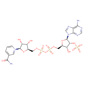
| HET Code: | NAP |
|---|---|
| Formula: | C21H25N7O17P3 |
| Molecular weight: | 740.381 g/mol |
| DrugBank ID: | DB03461 |
| Buried Surface Area: | 73.22 % |
| Polar Surface area: | 405.54 Å2 |
| Number of | |
|---|---|
| H-Bond Acceptors: | 21 |
| H-Bond Donors: | 5 |
| Rings: | 5 |
| Aromatic rings: | 3 |
| Anionic atoms: | 4 |
| Cationic atoms: | 1 |
| Rule of Five Violation: | 2 |
| Rotatable Bonds: | 13 |
| X | Y | Z |
|---|---|---|
| -37.8185 | -23.328 | 4.26367 |
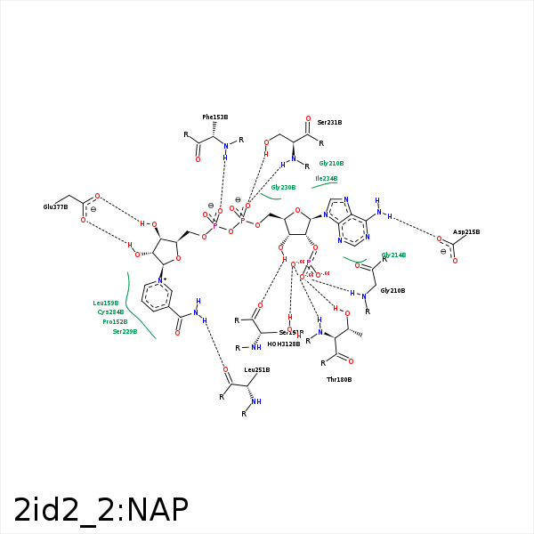


Represent the protein/ligand binding mode, centered on the ligand
Dashed lines represents hydrogen bonds and metal interactions
Green residue labels for amino acids with hydrophobic contacts (green lines) to the ligand
| Ligand | Protein | Interaction | |||
|---|---|---|---|---|---|
| Atom | Atom | Residue | Distance (Å) | Angle (°) | Type |
| C1B | CG2 | ILE- 150 | 3.97 | 0 | Hydrophobic |
| C4B | CG2 | ILE- 150 | 3.68 | 0 | Hydrophobic |
| O3B | O | SER- 151 | 2.91 | 174.67 | H-Bond (Ligand Donor) |
| C5N | CG | PRO- 152 | 3.3 | 0 | Hydrophobic |
| O2N | N | PHE- 153 | 3.24 | 165.62 | H-Bond (Protein Donor) |
| C5D | CE2 | PHE- 153 | 3.9 | 0 | Hydrophobic |
| C4N | CD2 | LEU- 159 | 3.45 | 0 | Hydrophobic |
| O2X | NZ | LYS- 177 | 3.36 | 147.64 | H-Bond (Protein Donor) |
| O2X | NZ | LYS- 177 | 3.36 | 0 | Ionic (Protein Cationic) |
| C3B | CB | PRO- 179 | 3.87 | 0 | Hydrophobic |
| O1X | N | THR- 180 | 2.59 | 159.36 | H-Bond (Protein Donor) |
| O2X | OG1 | THR- 180 | 2.83 | 147.97 | H-Bond (Protein Donor) |
| O2X | CZ | ARG- 209 | 3.74 | 0 | Ionic (Protein Cationic) |
| O3X | CZ | ARG- 209 | 3.4 | 0 | Ionic (Protein Cationic) |
| O2X | N | GLY- 210 | 2.73 | 153.72 | H-Bond (Protein Donor) |
| N6A | OD2 | ASP- 215 | 2.88 | 157.78 | H-Bond (Ligand Donor) |
| C1B | CE1 | PHE- 228 | 4.21 | 0 | Hydrophobic |
| C4B | CE1 | PHE- 228 | 3.63 | 0 | Hydrophobic |
| C4N | CB | SER- 229 | 4 | 0 | Hydrophobic |
| O2A | N | SER- 231 | 2.62 | 164.97 | H-Bond (Protein Donor) |
| O2A | OG | SER- 231 | 2.58 | 167.53 | H-Bond (Protein Donor) |
| O3 | N | SER- 231 | 3.3 | 127.66 | H-Bond (Protein Donor) |
| C4D | CB | SER- 231 | 4.36 | 0 | Hydrophobic |
| C3N | CB | GLU- 250 | 3.91 | 0 | Hydrophobic |
| N7N | O | LEU- 251 | 2.98 | 173.63 | H-Bond (Ligand Donor) |
| C2D | SG | CYS- 284 | 3.68 | 0 | Hydrophobic |
| C5N | SG | CYS- 284 | 3.24 | 0 | Hydrophobic |
| O3D | OE1 | GLU- 377 | 2.82 | 159.85 | H-Bond (Ligand Donor) |
| O2D | OE2 | GLU- 377 | 3.04 | 141.65 | H-Bond (Ligand Donor) |
| O2D | OE1 | GLU- 377 | 3.4 | 152.68 | H-Bond (Ligand Donor) |
| C5D | CZ | PHE- 379 | 3.73 | 0 | Hydrophobic |
| C2D | CE1 | PHE- 379 | 3.57 | 0 | Hydrophobic |
| O1X | O | HOH- 3128 | 2.88 | 179.97 | H-Bond (Protein Donor) |
