sc-PDB
An Annotated Database of Druggable Binding Sites from the Protein DataBank
An Annotated Database of Druggable Binding Sites from the Protein DataBank
1.580 Å
X-ray
2006-07-29
| Min | Mean | Median | Standard Deviation | Max | Count | |
|---|---|---|---|---|---|---|
| pChEMBL: | 7.720 | 7.720 | 7.720 | 0.000 | 7.720 | 1 |
| Name: | Aldose reductase |
|---|---|
| ID: | ALDR_HUMAN |
| AC: | P15121 |
| Organism: | Homo sapiens |
| Reign: | Eukaryota |
| TaxID: | 9606 |
| EC Number: | 1.1.1.21 |
| Chain Name: | Percentage of Residues within binding site |
|---|---|
| A | 100 % |
| B-Factor: | 15.011 |
|---|---|
| Number of residues: | 33 |
| Including | |
| Standard Amino Acids: | 32 |
| Non Standard Amino Acids: | 1 |
| Water Molecules: | 0 |
| Cofactors: | NAP |
| Metals: | |
| Ligandability | Volume (Å3) |
|---|---|
| 0.774 | 560.250 |
| % Hydrophobic | % Polar |
|---|---|
| 53.01 | 46.99 |
| According to VolSite | |
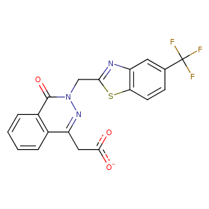
| HET Code: | ZST |
|---|---|
| Formula: | C19H11F3N3O3S |
| Molecular weight: | 418.369 g/mol |
| DrugBank ID: | DB08772 |
| Buried Surface Area: | 78.16 % |
| Polar Surface area: | 113.93 Å2 |
| Number of | |
|---|---|
| H-Bond Acceptors: | 5 |
| H-Bond Donors: | 0 |
| Rings: | 4 |
| Aromatic rings: | 3 |
| Anionic atoms: | 1 |
| Cationic atoms: | 0 |
| Rule of Five Violation: | 0 |
| Rotatable Bonds: | 5 |
| X | Y | Z |
|---|---|---|
| 16.4465 | -6.38169 | 14.1477 |
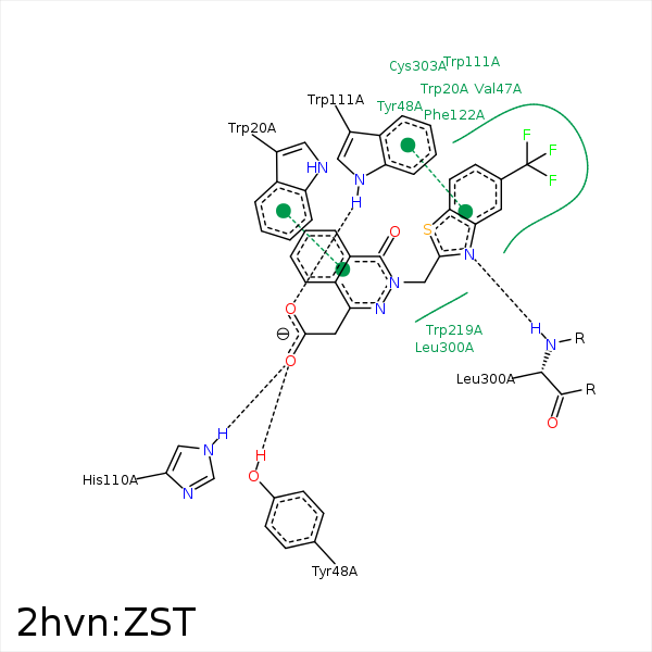


Represent the protein/ligand binding mode, centered on the ligand
Dashed lines represents hydrogen bonds and metal interactions
Green residue labels for amino acids with hydrophobic contacts (green lines) to the ligand
| Ligand | Protein | Interaction | |||
|---|---|---|---|---|---|
| Atom | Atom | Residue | Distance (Å) | Angle (°) | Type |
| C17 | CD2 | TRP- 20 | 3.75 | 0 | Hydrophobic |
| C7 | CG1 | VAL- 47 | 4.13 | 0 | Hydrophobic |
| C17 | CE1 | TYR- 48 | 4.29 | 0 | Hydrophobic |
| O3 | OH | TYR- 48 | 2.66 | 158.32 | H-Bond (Protein Donor) |
| S1 | CH2 | TRP- 79 | 4.09 | 0 | Hydrophobic |
| C13 | SG | CYS- 80 | 4.37 | 0 | Hydrophobic |
| O3 | NE2 | HIS- 110 | 2.7 | 152.5 | H-Bond (Protein Donor) |
| S1 | CZ2 | TRP- 111 | 3.71 | 0 | Hydrophobic |
| C14 | CB | TRP- 111 | 3.81 | 0 | Hydrophobic |
| F1 | CE3 | TRP- 111 | 3.21 | 0 | Hydrophobic |
| O2 | NE1 | TRP- 111 | 2.94 | 163.3 | H-Bond (Protein Donor) |
| DuAr | DuAr | TRP- 111 | 3.38 | 0 | Aromatic Face/Face |
| F2 | CG2 | THR- 113 | 3.3 | 0 | Hydrophobic |
| S1 | CZ | PHE- 122 | 3.82 | 0 | Hydrophobic |
| C9 | CH2 | TRP- 219 | 3.54 | 0 | Hydrophobic |
| C9 | CB | CYS- 298 | 4.14 | 0 | Hydrophobic |
| C17 | SG | CYS- 298 | 4.19 | 0 | Hydrophobic |
| C9 | CB | LEU- 300 | 4.09 | 0 | Hydrophobic |
| C16 | CB | LEU- 300 | 4.41 | 0 | Hydrophobic |
| N3 | N | LEU- 300 | 3.18 | 154.82 | H-Bond (Protein Donor) |
| C14 | SG | CYS- 303 | 4.18 | 0 | Hydrophobic |
| C15 | CB | CYS- 303 | 4.01 | 0 | Hydrophobic |
| F2 | CB | CYS- 303 | 3.22 | 0 | Hydrophobic |
| F3 | CG | TYR- 309 | 4.12 | 0 | Hydrophobic |
| F2 | CD1 | TYR- 309 | 3.42 | 0 | Hydrophobic |
| F1 | CG | PRO- 310 | 3.91 | 0 | Hydrophobic |
| F3 | CG | PRO- 310 | 3.69 | 0 | Hydrophobic |
| C17 | C4N | NAP- 500 | 3.51 | 0 | Hydrophobic |
