sc-PDB
An Annotated Database of Druggable Binding Sites from the Protein DataBank
An Annotated Database of Druggable Binding Sites from the Protein DataBank
1.700 Å
X-ray
2006-03-25
| Name: | Glutathione reductase, mitochondrial |
|---|---|
| ID: | GSHR_HUMAN |
| AC: | P00390 |
| Organism: | Homo sapiens |
| Reign: | Eukaryota |
| TaxID: | 9606 |
| EC Number: | 1.8.1.7 |
| Chain Name: | Percentage of Residues within binding site |
|---|---|
| A | 94 % |
| B | 6 % |
| B-Factor: | 25.521 |
|---|---|
| Number of residues: | 75 |
| Including | |
| Standard Amino Acids: | 66 |
| Non Standard Amino Acids: | 1 |
| Water Molecules: | 8 |
| Cofactors: | |
| Metals: | |
| Ligandability | Volume (Å3) |
|---|---|
| 1.237 | 1164.375 |
| % Hydrophobic | % Polar |
|---|---|
| 40.87 | 59.13 |
| According to VolSite | |
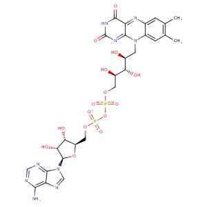
| HET Code: | FAD |
|---|---|
| Formula: | C27H31N9O15P2 |
| Molecular weight: | 783.534 g/mol |
| DrugBank ID: | DB03147 |
| Buried Surface Area: | 73.65 % |
| Polar Surface area: | 381.7 Å2 |
| Number of | |
|---|---|
| H-Bond Acceptors: | 22 |
| H-Bond Donors: | 7 |
| Rings: | 6 |
| Aromatic rings: | 3 |
| Anionic atoms: | 2 |
| Cationic atoms: | 0 |
| Rule of Five Violation: | 3 |
| Rotatable Bonds: | 13 |
| X | Y | Z |
|---|---|---|
| 92.3548 | 18.0727 | 50.6559 |


Represent the protein/ligand binding mode, centered on the ligand
Dashed lines represents hydrogen bonds and metal interactions
Green residue labels for amino acids with hydrophobic contacts (green lines) to the ligand
| Ligand | Protein | Interaction | |||
|---|---|---|---|---|---|
| Atom | Atom | Residue | Distance (Å) | Angle (°) | Type |
| O1P | N | GLY- 31 | 2.86 | 166.51 | H-Bond (Protein Donor) |
| O3B | OE2 | GLU- 50 | 3.16 | 126.32 | H-Bond (Ligand Donor) |
| O3B | OE1 | GLU- 50 | 2.89 | 174.78 | H-Bond (Ligand Donor) |
| O2B | OE2 | GLU- 50 | 2.72 | 147.44 | H-Bond (Ligand Donor) |
| N3A | N | SER- 51 | 3.28 | 138.03 | H-Bond (Protein Donor) |
| O2B | ND1 | HIS- 52 | 3.15 | 164.65 | H-Bond (Protein Donor) |
| O1A | N | THR- 57 | 3.04 | 148.72 | H-Bond (Protein Donor) |
| O2A | OG1 | THR- 57 | 2.72 | 169.14 | H-Bond (Protein Donor) |
| C8M | CG2 | THR- 57 | 3.9 | 0 | Hydrophobic |
| C9A | SG | CYS- 63 | 4.36 | 0 | Hydrophobic |
| C2' | SG | CYS- 63 | 3.88 | 0 | Hydrophobic |
| O4 | NZ | LYS- 66 | 2.79 | 134.57 | H-Bond (Protein Donor) |
| N5 | NZ | LYS- 66 | 2.98 | 134.77 | H-Bond (Protein Donor) |
| C6 | CG | LYS- 66 | 4.45 | 0 | Hydrophobic |
| N6A | O | ALA- 130 | 3.1 | 152.01 | H-Bond (Ligand Donor) |
| N1A | N | ALA- 130 | 3.01 | 163.82 | H-Bond (Protein Donor) |
| C7M | CB | SER- 177 | 3.9 | 0 | Hydrophobic |
| C7M | CE2 | PHE- 181 | 4.15 | 0 | Hydrophobic |
| C7M | CG2 | ILE- 198 | 4.27 | 0 | Hydrophobic |
| C8M | CD1 | ILE- 198 | 3.87 | 0 | Hydrophobic |
| C8 | CD1 | ILE- 198 | 3.81 | 0 | Hydrophobic |
| O3' | OD2 | ASP- 331 | 3.49 | 133.17 | H-Bond (Ligand Donor) |
| O3' | OD1 | ASP- 331 | 2.79 | 172.05 | H-Bond (Ligand Donor) |
| C5' | CB | ASP- 331 | 4.23 | 0 | Hydrophobic |
| O2P | N | ASP- 331 | 2.87 | 150.75 | H-Bond (Protein Donor) |
| N1 | N | THR- 339 | 3.41 | 152.48 | H-Bond (Protein Donor) |
| O2 | N | THR- 339 | 2.99 | 145.21 | H-Bond (Protein Donor) |
| C2' | CB | THR- 339 | 4.4 | 0 | Hydrophobic |
| N3 | O | HIS- 467 | 2.73 | 155.33 | H-Bond (Ligand Donor) |
| O1P | O | HOH- 5707 | 2.8 | 170.4 | H-Bond (Protein Donor) |
| O2P | O | HOH- 5713 | 2.68 | 179.97 | H-Bond (Protein Donor) |
| O2A | O | HOH- 5821 | 2.98 | 138.37 | H-Bond (Protein Donor) |
