sc-PDB
An Annotated Database of Druggable Binding Sites from the Protein DataBank
An Annotated Database of Druggable Binding Sites from the Protein DataBank
2.580 Å
X-ray
2005-07-26
| Name: | Benzaldehyde lyase |
|---|---|
| ID: | Q9F4L3_PSEFL |
| AC: | Q9F4L3 |
| Organism: | Pseudomonas fluorescens |
| Reign: | Bacteria |
| TaxID: | 294 |
| EC Number: | / |
| Chain Name: | Percentage of Residues within binding site |
|---|---|
| A | 71 % |
| B | 29 % |
| B-Factor: | 44.615 |
|---|---|
| Number of residues: | 42 |
| Including | |
| Standard Amino Acids: | 40 |
| Non Standard Amino Acids: | 1 |
| Water Molecules: | 1 |
| Cofactors: | |
| Metals: | MG |
| Ligandability | Volume (Å3) |
|---|---|
| 0.656 | 330.750 |
| % Hydrophobic | % Polar |
|---|---|
| 51.02 | 48.98 |
| According to VolSite | |
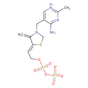
| HET Code: | TPP |
|---|---|
| Formula: | C12H16N4O7P2S |
| Molecular weight: | 422.291 g/mol |
| DrugBank ID: | - |
| Buried Surface Area: | 79.98 % |
| Polar Surface area: | 225.32 Å2 |
| Number of | |
|---|---|
| H-Bond Acceptors: | 10 |
| H-Bond Donors: | 1 |
| Rings: | 2 |
| Aromatic rings: | 2 |
| Anionic atoms: | 3 |
| Cationic atoms: | 1 |
| Rule of Five Violation: | 1 |
| Rotatable Bonds: | 8 |
| X | Y | Z |
|---|---|---|
| 41.6127 | -26.7498 | 6.74812 |
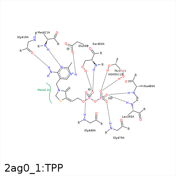


Represent the protein/ligand binding mode, centered on the ligand
Dashed lines represents hydrogen bonds and metal interactions
Green residue labels for amino acids with hydrophobic contacts (green lines) to the ligand
| Ligand | Protein | Interaction | |||
|---|---|---|---|---|---|
| Atom | Atom | Residue | Distance (Å) | Angle (°) | Type |
| N1' | OE2 | GLU- 50 | 2.86 | 152.46 | H-Bond (Ligand Donor) |
| C5' | CG2 | THR- 73 | 3.99 | 0 | Hydrophobic |
| S1 | CB | ALA- 394 | 3.7 | 0 | Hydrophobic |
| C7 | CB | ALA- 394 | 3.84 | 0 | Hydrophobic |
| O2B | N | LEU- 395 | 3.21 | 149.97 | H-Bond (Protein Donor) |
| O1B | OG1 | THR- 396 | 3.03 | 160.05 | H-Bond (Protein Donor) |
| O1B | N | THR- 396 | 3.41 | 137.68 | H-Bond (Protein Donor) |
| N4' | O | GLY- 419 | 2.93 | 168.28 | H-Bond (Ligand Donor) |
| CM2 | CB | SER- 420 | 4.07 | 0 | Hydrophobic |
| N3' | N | MET- 421 | 3.33 | 159.12 | H-Bond (Protein Donor) |
| C5' | CG | MET- 421 | 4.23 | 0 | Hydrophobic |
| C7 | SD | MET- 421 | 4.19 | 0 | Hydrophobic |
| S1 | CE | MET- 421 | 3.83 | 0 | Hydrophobic |
| O1A | N | GLY- 449 | 2.73 | 149.94 | H-Bond (Protein Donor) |
| O2A | N | SER- 450 | 2.83 | 141.76 | H-Bond (Protein Donor) |
| O2A | OG | SER- 450 | 2.67 | 163.66 | H-Bond (Protein Donor) |
| CM2 | CZ | TYR- 453 | 4.1 | 0 | Hydrophobic |
| O3B | ND2 | ASN- 475 | 3.33 | 137.85 | H-Bond (Protein Donor) |
| C7 | CE3 | TRP- 478 | 4.15 | 0 | Hydrophobic |
| O3B | N | GLY- 479 | 2.83 | 139.9 | H-Bond (Protein Donor) |
| S1 | CB | ALA- 480 | 4.13 | 0 | Hydrophobic |
| O2B | N | ALA- 480 | 3.22 | 150.67 | H-Bond (Protein Donor) |
| C7' | CG2 | THR- 481 | 4.18 | 0 | Hydrophobic |
| C2 | CG2 | THR- 481 | 3.61 | 0 | Hydrophobic |
| O1A | MG | MG- 601 | 2.21 | 0 | Metal Acceptor |
| O3B | MG | MG- 601 | 2.01 | 0 | Metal Acceptor |
| O1B | O | HOH- 5011 | 2.7 | 120.26 | H-Bond (Protein Donor) |
