sc-PDB
An Annotated Database of Druggable Binding Sites from the Protein DataBank
An Annotated Database of Druggable Binding Sites from the Protein DataBank
1.760 Å
X-ray
1994-04-15
| Name: | Aldose reductase |
|---|---|
| ID: | ALDR_HUMAN |
| AC: | P15121 |
| Organism: | Homo sapiens |
| Reign: | Eukaryota |
| TaxID: | 9606 |
| EC Number: | 1.1.1.21 |
| Chain Name: | Percentage of Residues within binding site |
|---|---|
| A | 100 % |
| B-Factor: | 9.065 |
|---|---|
| Number of residues: | 47 |
| Including | |
| Standard Amino Acids: | 45 |
| Non Standard Amino Acids: | 0 |
| Water Molecules: | 2 |
| Cofactors: | |
| Metals: | |
| Ligandability | Volume (Å3) |
|---|---|
| 0.742 | 816.750 |
| % Hydrophobic | % Polar |
|---|---|
| 49.59 | 50.41 |
| According to VolSite | |
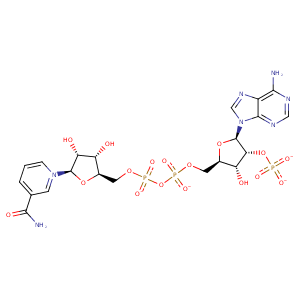
| HET Code: | NAP |
|---|---|
| Formula: | C21H25N7O17P3 |
| Molecular weight: | 740.381 g/mol |
| DrugBank ID: | DB03461 |
| Buried Surface Area: | 78.55 % |
| Polar Surface area: | 405.54 Å2 |
| Number of | |
|---|---|
| H-Bond Acceptors: | 21 |
| H-Bond Donors: | 5 |
| Rings: | 5 |
| Aromatic rings: | 3 |
| Anionic atoms: | 4 |
| Cationic atoms: | 1 |
| Rule of Five Violation: | 2 |
| Rotatable Bonds: | 13 |
| X | Y | Z |
|---|---|---|
| 22.1515 | 26.9256 | 72.9159 |


Represent the protein/ligand binding mode, centered on the ligand
Dashed lines represents hydrogen bonds and metal interactions
Green residue labels for amino acids with hydrophobic contacts (green lines) to the ligand
| Ligand | Protein | Interaction | |||
|---|---|---|---|---|---|
| Atom | Atom | Residue | Distance (Å) | Angle (°) | Type |
| O2D | N | THR- 19 | 3.32 | 146.85 | H-Bond (Protein Donor) |
| O3D | N | TRP- 20 | 2.94 | 144.92 | H-Bond (Protein Donor) |
| C3D | CB | TRP- 20 | 3.74 | 0 | Hydrophobic |
| O1N | NZ | LYS- 21 | 2.84 | 149.44 | H-Bond (Protein Donor) |
| O1N | NZ | LYS- 21 | 2.84 | 0 | Ionic (Protein Cationic) |
| O2D | OD2 | ASP- 43 | 2.74 | 150.78 | H-Bond (Ligand Donor) |
| C2D | CZ | TYR- 48 | 4.13 | 0 | Hydrophobic |
| N7N | OG | SER- 159 | 2.89 | 140.85 | H-Bond (Ligand Donor) |
| O7N | ND2 | ASN- 160 | 3.1 | 164.03 | H-Bond (Protein Donor) |
| N7N | OE1 | GLN- 183 | 3.04 | 158.07 | H-Bond (Ligand Donor) |
| C3N | CB | TYR- 209 | 4.3 | 0 | Hydrophobic |
| DuAr | DuAr | TYR- 209 | 3.55 | 0 | Aromatic Face/Face |
| O2N | OG | SER- 210 | 2.7 | 152.01 | H-Bond (Protein Donor) |
| O5D | N | SER- 210 | 3.12 | 133.77 | H-Bond (Protein Donor) |
| O1A | N | LEU- 212 | 3.11 | 142.4 | H-Bond (Protein Donor) |
| C1B | CD1 | LEU- 212 | 4.29 | 0 | Hydrophobic |
| O1A | N | SER- 214 | 3.06 | 142.97 | H-Bond (Protein Donor) |
| O2N | OG | SER- 214 | 2.8 | 150.34 | H-Bond (Protein Donor) |
| C4B | CG | PRO- 215 | 3.58 | 0 | Hydrophobic |
| C1B | CG | PRO- 215 | 4.17 | 0 | Hydrophobic |
| C3B | CB | ASP- 216 | 4.27 | 0 | Hydrophobic |
| C4D | CG1 | ILE- 260 | 4.19 | 0 | Hydrophobic |
| O2A | N | LYS- 262 | 2.88 | 172.32 | H-Bond (Protein Donor) |
| O1X | NZ | LYS- 262 | 2.79 | 172.94 | H-Bond (Protein Donor) |
| C5B | CD | LYS- 262 | 3.88 | 0 | Hydrophobic |
| C3B | CD | LYS- 262 | 3.92 | 0 | Hydrophobic |
| C5D | CB | LYS- 262 | 3.93 | 0 | Hydrophobic |
| O1X | NZ | LYS- 262 | 2.79 | 0 | Ionic (Protein Cationic) |
| O3X | OG | SER- 263 | 2.8 | 167.82 | H-Bond (Protein Donor) |
| O1X | N | VAL- 264 | 3.07 | 155.22 | H-Bond (Protein Donor) |
| O3X | OG1 | THR- 265 | 2.82 | 158.31 | H-Bond (Protein Donor) |
| O3X | CZ | ARG- 268 | 3.85 | 0 | Ionic (Protein Cationic) |
| N6A | OE2 | GLU- 271 | 3.07 | 167.03 | H-Bond (Ligand Donor) |
| N7A | ND2 | ASN- 272 | 3.06 | 170.99 | H-Bond (Protein Donor) |
| N6A | OD1 | ASN- 272 | 2.89 | 144.83 | H-Bond (Ligand Donor) |
| C4N | SG | CYS- 298 | 4.14 | 0 | Hydrophobic |
| N1A | O | HOH- 917 | 2.93 | 179.94 | H-Bond (Protein Donor) |
