sc-PDB
An Annotated Database of Druggable Binding Sites from the Protein DataBank
An Annotated Database of Druggable Binding Sites from the Protein DataBank
2.200 Å
X-ray
2005-02-08
| Name: | Uncharacterized protein |
|---|---|
| ID: | O59611_PYRHO |
| AC: | O59611 |
| Organism: | Pyrococcus horikoshii |
| Reign: | Archaea |
| TaxID: | 70601 |
| EC Number: | / |
| Chain Name: | Percentage of Residues within binding site |
|---|---|
| C | 100 % |
| B-Factor: | 46.709 |
|---|---|
| Number of residues: | 37 |
| Including | |
| Standard Amino Acids: | 36 |
| Non Standard Amino Acids: | 0 |
| Water Molecules: | 1 |
| Cofactors: | |
| Metals: | |
| Ligandability | Volume (Å3) |
|---|---|
| 0.201 | 378.000 |
| % Hydrophobic | % Polar |
|---|---|
| 42.86 | 57.14 |
| According to VolSite | |
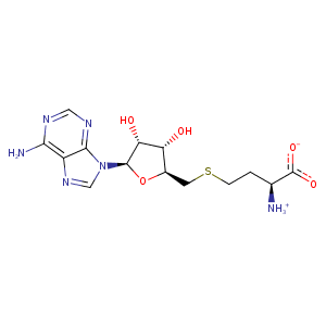
| HET Code: | SAH |
|---|---|
| Formula: | C14H20N6O5S |
| Molecular weight: | 384.411 g/mol |
| DrugBank ID: | DB01752 |
| Buried Surface Area: | 78.8 % |
| Polar Surface area: | 212.38 Å2 |
| Number of | |
|---|---|
| H-Bond Acceptors: | 10 |
| H-Bond Donors: | 4 |
| Rings: | 3 |
| Aromatic rings: | 2 |
| Anionic atoms: | 1 |
| Cationic atoms: | 1 |
| Rule of Five Violation: | 1 |
| Rotatable Bonds: | 7 |
| X | Y | Z |
|---|---|---|
| 12.2121 | 7.14658 | 6.88219 |
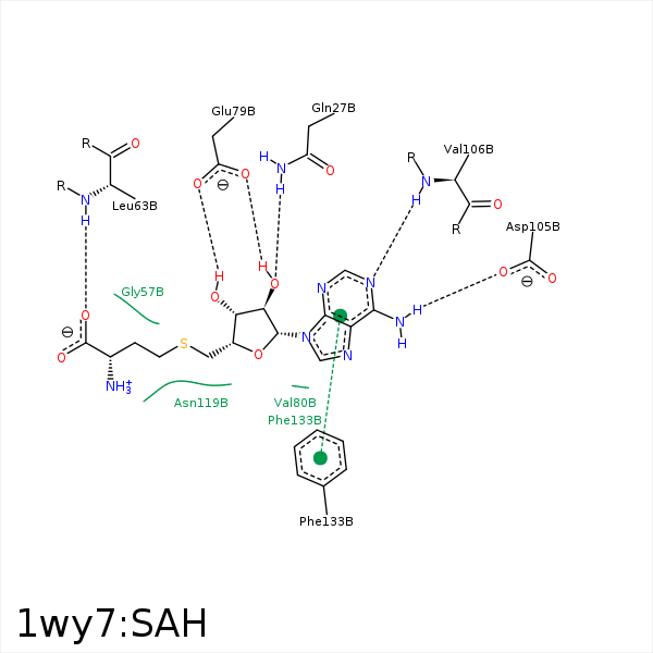


Represent the protein/ligand binding mode, centered on the ligand
Dashed lines represents hydrogen bonds and metal interactions
Green residue labels for amino acids with hydrophobic contacts (green lines) to the ligand
| Ligand | Protein | Interaction | |||
|---|---|---|---|---|---|
| Atom | Atom | Residue | Distance (Å) | Angle (°) | Type |
| CB | CZ | PHE- 18 | 4.11 | 0 | Hydrophobic |
| C3' | CZ | PHE- 18 | 4.32 | 0 | Hydrophobic |
| C2' | CB | LEU- 25 | 4.25 | 0 | Hydrophobic |
| O2' | NE2 | GLN- 27 | 2.99 | 168.65 | H-Bond (Protein Donor) |
| C3' | CG | GLN- 27 | 3.85 | 0 | Hydrophobic |
| O | N | LEU- 63 | 3.27 | 175.62 | H-Bond (Protein Donor) |
| O3' | OE1 | GLU- 79 | 2.96 | 175.35 | H-Bond (Ligand Donor) |
| O2' | OE2 | GLU- 79 | 2.66 | 151.7 | H-Bond (Ligand Donor) |
| O2' | OE1 | GLU- 79 | 3.42 | 134.66 | H-Bond (Ligand Donor) |
| N3 | N | VAL- 80 | 3.48 | 146.06 | H-Bond (Protein Donor) |
| N6 | OD2 | ASP- 105 | 2.91 | 148.56 | H-Bond (Ligand Donor) |
| N1 | N | VAL- 106 | 3.1 | 148.87 | H-Bond (Protein Donor) |
| CG | CB | ASN- 119 | 4.32 | 0 | Hydrophobic |
