sc-PDB
An Annotated Database of Druggable Binding Sites from the Protein DataBank
An Annotated Database of Druggable Binding Sites from the Protein DataBank
2.650 Å
X-ray
2004-07-27
| Name: | L-lactate dehydrogenase |
|---|---|
| ID: | LDH_PLAFD |
| AC: | Q27743 |
| Organism: | Plasmodium falciparum |
| Reign: | Eukaryota |
| TaxID: | 5836 |
| EC Number: | 1.1.1.27 |
| Chain Name: | Percentage of Residues within binding site |
|---|---|
| A | 100 % |
| B-Factor: | 20.605 |
|---|---|
| Number of residues: | 21 |
| Including | |
| Standard Amino Acids: | 20 |
| Non Standard Amino Acids: | 1 |
| Water Molecules: | 0 |
| Cofactors: | NAD |
| Metals: | |
| Ligandability | Volume (Å3) |
|---|---|
| 0.220 | 330.750 |
| % Hydrophobic | % Polar |
|---|---|
| 39.80 | 60.20 |
| According to VolSite | |
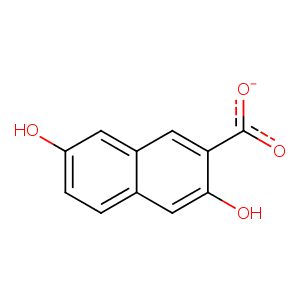
| HET Code: | BIK |
|---|---|
| Formula: | C11H7O4 |
| Molecular weight: | 203.171 g/mol |
| DrugBank ID: | DB04641 |
| Buried Surface Area: | 46.08 % |
| Polar Surface area: | 80.59 Å2 |
| Number of | |
|---|---|
| H-Bond Acceptors: | 4 |
| H-Bond Donors: | 2 |
| Rings: | 2 |
| Aromatic rings: | 2 |
| Anionic atoms: | 1 |
| Cationic atoms: | 0 |
| Rule of Five Violation: | 0 |
| Rotatable Bonds: | 1 |
| X | Y | Z |
|---|---|---|
| 23.6663 | 17.8447 | 5.09333 |
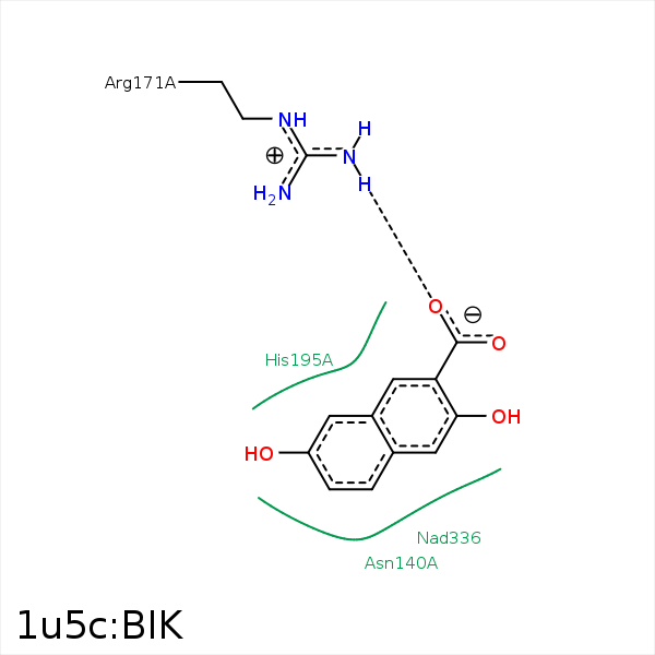


Represent the protein/ligand binding mode, centered on the ligand
Dashed lines represents hydrogen bonds and metal interactions
Green residue labels for amino acids with hydrophobic contacts (green lines) to the ligand
| Ligand | Protein | Interaction | |||
|---|---|---|---|---|---|
| Atom | Atom | Residue | Distance (Å) | Angle (°) | Type |
| C7 | CB | ASN- 140 | 3.49 | 0 | Hydrophobic |
| O32 | CZ | ARG- 171 | 3.95 | 0 | Ionic (Protein Cationic) |
| O32 | NH2 | ARG- 171 | 3.07 | 175.22 | H-Bond (Protein Donor) |
| O31 | NH1 | ARG- 171 | 3.41 | 170.44 | H-Bond (Protein Donor) |
| C3 | CB | ALA- 236 | 3.69 | 0 | Hydrophobic |
