sc-PDB
An Annotated Database of Druggable Binding Sites from the Protein DataBank
An Annotated Database of Druggable Binding Sites from the Protein DataBank
1.800 Å
X-ray
2003-12-22
| Name: | Glycine oxidase |
|---|---|
| ID: | GLOX_BACSU |
| AC: | O31616 |
| Organism: | Bacillus subtilis |
| Reign: | Bacteria |
| TaxID: | 224308 |
| EC Number: | 1.4.3.19 |
| Chain Name: | Percentage of Residues within binding site |
|---|---|
| C | 100 % |
| B-Factor: | 18.989 |
|---|---|
| Number of residues: | 70 |
| Including | |
| Standard Amino Acids: | 66 |
| Non Standard Amino Acids: | 0 |
| Water Molecules: | 4 |
| Cofactors: | |
| Metals: | |
| Ligandability | Volume (Å3) |
|---|---|
| 1.027 | 752.625 |
| % Hydrophobic | % Polar |
|---|---|
| 44.39 | 55.61 |
| According to VolSite | |
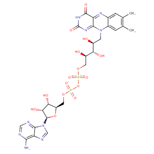
| HET Code: | FAD |
|---|---|
| Formula: | C27H31N9O15P2 |
| Molecular weight: | 783.534 g/mol |
| DrugBank ID: | DB03147 |
| Buried Surface Area: | 76.96 % |
| Polar Surface area: | 381.7 Å2 |
| Number of | |
|---|---|
| H-Bond Acceptors: | 22 |
| H-Bond Donors: | 7 |
| Rings: | 6 |
| Aromatic rings: | 3 |
| Anionic atoms: | 2 |
| Cationic atoms: | 0 |
| Rule of Five Violation: | 3 |
| Rotatable Bonds: | 13 |
| X | Y | Z |
|---|---|---|
| 11.7262 | 185.736 | 156.805 |


Represent the protein/ligand binding mode, centered on the ligand
Dashed lines represents hydrogen bonds and metal interactions
Green residue labels for amino acids with hydrophobic contacts (green lines) to the ligand
| Ligand | Protein | Interaction | |||
|---|---|---|---|---|---|
| Atom | Atom | Residue | Distance (Å) | Angle (°) | Type |
| C5' | CG1 | ILE- 15 | 4.43 | 0 | Hydrophobic |
| O2P | N | ILE- 15 | 3.24 | 158.32 | H-Bond (Protein Donor) |
| O3B | OE2 | GLU- 34 | 3.19 | 121.96 | H-Bond (Ligand Donor) |
| O3B | OE1 | GLU- 34 | 2.61 | 171.55 | H-Bond (Ligand Donor) |
| O2B | OE2 | GLU- 34 | 2.66 | 164.5 | H-Bond (Ligand Donor) |
| N3A | N | SER- 35 | 3.25 | 146.63 | H-Bond (Protein Donor) |
| O2B | NH1 | ARG- 41 | 3.49 | 135.16 | H-Bond (Protein Donor) |
| C3B | CG | ARG- 41 | 3.47 | 0 | Hydrophobic |
| O2A | N | THR- 42 | 3.36 | 151.05 | H-Bond (Protein Donor) |
| C8M | CG2 | THR- 42 | 4.07 | 0 | Hydrophobic |
| C9 | CG2 | THR- 42 | 4.2 | 0 | Hydrophobic |
| C3' | CG2 | THR- 42 | 4.21 | 0 | Hydrophobic |
| O1A | N | THR- 43 | 3 | 149.36 | H-Bond (Protein Donor) |
| O1A | OG1 | THR- 43 | 2.7 | 157.84 | H-Bond (Protein Donor) |
| O4' | OG1 | THR- 43 | 3.37 | 156.62 | H-Bond (Ligand Donor) |
| C8M | CB | ALA- 45 | 4.47 | 0 | Hydrophobic |
| C6 | CB | ALA- 46 | 4.28 | 0 | Hydrophobic |
| C2' | CB | ALA- 46 | 4.16 | 0 | Hydrophobic |
| C9A | CB | ALA- 46 | 3.65 | 0 | Hydrophobic |
| N5 | N | ALA- 47 | 3.29 | 151.21 | H-Bond (Protein Donor) |
| O4 | N | GLY- 48 | 3 | 132.21 | H-Bond (Protein Donor) |
| N3 | O | MET- 49 | 2.98 | 169.67 | H-Bond (Ligand Donor) |
| O4 | N | MET- 49 | 2.87 | 138.4 | H-Bond (Protein Donor) |
| N6A | O | VAL- 174 | 2.94 | 168.69 | H-Bond (Ligand Donor) |
| N1A | N | VAL- 174 | 2.99 | 177.03 | H-Bond (Protein Donor) |
| C3B | CZ3 | TRP- 205 | 4.4 | 0 | Hydrophobic |
| C2B | CE3 | TRP- 205 | 4.24 | 0 | Hydrophobic |
| C7M | SG | CYS- 226 | 3.59 | 0 | Hydrophobic |
| C7M | CB | ALA- 259 | 4.08 | 0 | Hydrophobic |
| C8M | CG | ARG- 302 | 3.63 | 0 | Hydrophobic |
| O3' | O | HIS- 327 | 2.62 | 156.2 | H-Bond (Ligand Donor) |
| O3' | N | GLY- 331 | 2.99 | 122.53 | H-Bond (Protein Donor) |
| O2 | N | ILE- 332 | 3.01 | 127.56 | H-Bond (Protein Donor) |
| C2' | CD1 | ILE- 332 | 4.25 | 0 | Hydrophobic |
| C4' | CD1 | ILE- 332 | 4.37 | 0 | Hydrophobic |
| O2 | N | LEU- 333 | 3.07 | 169.72 | H-Bond (Protein Donor) |
| O1P | O | HOH- 9312 | 2.75 | 153.51 | H-Bond (Protein Donor) |
| O2P | O | HOH- 9313 | 2.67 | 179.95 | H-Bond (Protein Donor) |
| O1P | O | HOH- 9315 | 2.75 | 179.96 | H-Bond (Protein Donor) |
| O2A | O | HOH- 9316 | 2.95 | 141.84 | H-Bond (Protein Donor) |
