sc-PDB
An Annotated Database of Druggable Binding Sites from the Protein DataBank
An Annotated Database of Druggable Binding Sites from the Protein DataBank
2.100 Å
X-ray
1998-03-26
| Name: | Sepiapterin reductase |
|---|---|
| ID: | SPRE_MOUSE |
| AC: | Q64105 |
| Organism: | Mus musculus |
| Reign: | Eukaryota |
| TaxID: | 10090 |
| EC Number: | 1.1.1.153 |
| Chain Name: | Percentage of Residues within binding site |
|---|---|
| A | 100 % |
| B-Factor: | 18.904 |
|---|---|
| Number of residues: | 20 |
| Including | |
| Standard Amino Acids: | 19 |
| Non Standard Amino Acids: | 1 |
| Water Molecules: | 0 |
| Cofactors: | NAP |
| Metals: | |
| Ligandability | Volume (Å3) |
|---|---|
| 1.021 | 324.000 |
| % Hydrophobic | % Polar |
|---|---|
| 66.67 | 33.33 |
| According to VolSite | |
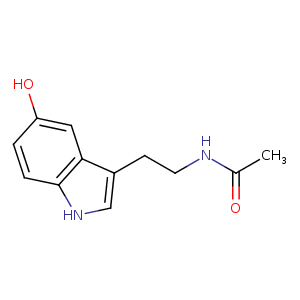
| HET Code: | ASE |
|---|---|
| Formula: | C12H14N2O2 |
| Molecular weight: | 218.252 g/mol |
| DrugBank ID: | DB04275 |
| Buried Surface Area: | 62.82 % |
| Polar Surface area: | 65.12 Å2 |
| Number of | |
|---|---|
| H-Bond Acceptors: | 2 |
| H-Bond Donors: | 3 |
| Rings: | 2 |
| Aromatic rings: | 2 |
| Anionic atoms: | 0 |
| Cationic atoms: | 0 |
| Rule of Five Violation: | 0 |
| Rotatable Bonds: | 3 |
| X | Y | Z |
|---|---|---|
| -15.9826 | 62.575 | 25.8931 |
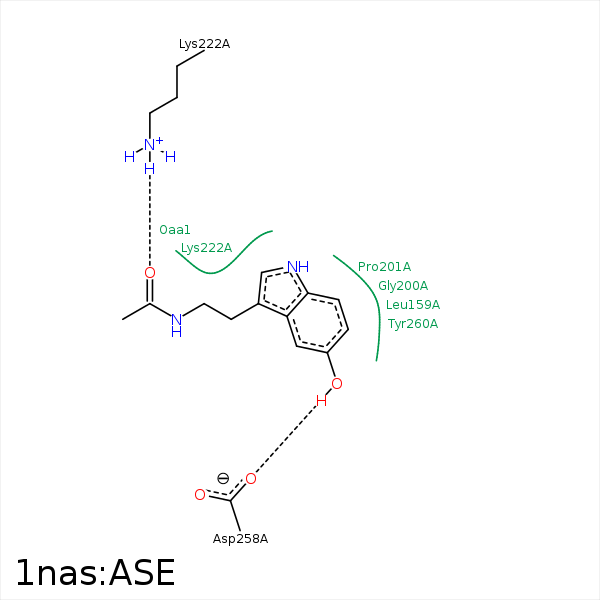


Represent the protein/ligand binding mode, centered on the ligand
Dashed lines represents hydrogen bonds and metal interactions
Green residue labels for amino acids with hydrophobic contacts (green lines) to the ligand
| Ligand | Protein | Interaction | |||
|---|---|---|---|---|---|
| Atom | Atom | Residue | Distance (Å) | Angle (°) | Type |
| C3 | CD2 | LEU- 159 | 3.77 | 0 | Hydrophobic |
| C1 | CB | LEU- 159 | 3.63 | 0 | Hydrophobic |
| C15 | CG | TYR- 165 | 3.37 | 0 | Hydrophobic |
| C15 | CH2 | TRP- 168 | 3.56 | 0 | Hydrophobic |
| O10 | N | GLY- 200 | 3.38 | 122.33 | H-Bond (Protein Donor) |
| C3 | CG | PRO- 201 | 4.34 | 0 | Hydrophobic |
| C11 | CB | PRO- 201 | 4.08 | 0 | Hydrophobic |
| C15 | CD1 | LEU- 219 | 3.38 | 0 | Hydrophobic |
| O10 | OD1 | ASP- 258 | 2.66 | 152.16 | H-Bond (Ligand Donor) |
