sc-PDB
An Annotated Database of Druggable Binding Sites from the Protein DataBank
An Annotated Database of Druggable Binding Sites from the Protein DataBank
1.600 Å
X-ray
2002-10-22
| Name: | Pantothenate synthetase |
|---|---|
| ID: | PANC_MYCTU |
| AC: | P9WIL5 |
| Organism: | Mycobacterium tuberculosis |
| Reign: | Bacteria |
| TaxID: | 83332 |
| EC Number: | 6.3.2.1 |
| Chain Name: | Percentage of Residues within binding site |
|---|---|
| B | 100 % |
| B-Factor: | 24.528 |
|---|---|
| Number of residues: | 41 |
| Including | |
| Standard Amino Acids: | 37 |
| Non Standard Amino Acids: | 1 |
| Water Molecules: | 3 |
| Cofactors: | |
| Metals: | MG |
| Ligandability | Volume (Å3) |
|---|---|
| 0.397 | 1042.875 |
| % Hydrophobic | % Polar |
|---|---|
| 38.51 | 61.49 |
| According to VolSite | |
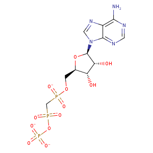
| HET Code: | APC |
|---|---|
| Formula: | C11H14N5O12P3 |
| Molecular weight: | 501.176 g/mol |
| DrugBank ID: | DB02596 |
| Buried Surface Area: | 73.63 % |
| Polar Surface area: | 310.64 Å2 |
| Number of | |
|---|---|
| H-Bond Acceptors: | 16 |
| H-Bond Donors: | 3 |
| Rings: | 3 |
| Aromatic rings: | 2 |
| Anionic atoms: | 4 |
| Cationic atoms: | 0 |
| Rule of Five Violation: | 2 |
| Rotatable Bonds: | 8 |
| X | Y | Z |
|---|---|---|
| 18.5198 | 13.1325 | 78.2305 |
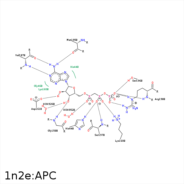


Represent the protein/ligand binding mode, centered on the ligand
Dashed lines represents hydrogen bonds and metal interactions
Green residue labels for amino acids with hydrophobic contacts (green lines) to the ligand
| Ligand | Protein | Interaction | |||
|---|---|---|---|---|---|
| Atom | Atom | Residue | Distance (Å) | Angle (°) | Type |
| O1B | NE2 | HIS- 44 | 2.79 | 169 | H-Bond (Protein Donor) |
| C1' | CD2 | LEU- 50 | 3.81 | 0 | Hydrophobic |
| C4' | CD2 | LEU- 50 | 3.9 | 0 | Hydrophobic |
| O3' | N | GLY- 158 | 3.03 | 164.69 | H-Bond (Protein Donor) |
| O2' | N | GLY- 158 | 3.31 | 124.46 | H-Bond (Protein Donor) |
| O1B | NZ | LYS- 160 | 3.54 | 0 | Ionic (Protein Cationic) |
| O2B | NZ | LYS- 160 | 2.82 | 0 | Ionic (Protein Cationic) |
| O2B | NZ | LYS- 160 | 2.82 | 143.95 | H-Bond (Protein Donor) |
| O2' | OD2 | ASP- 161 | 2.63 | 164.4 | H-Bond (Ligand Donor) |
| N6 | O | VAL- 187 | 2.96 | 160.68 | H-Bond (Ligand Donor) |
| N1 | N | VAL- 187 | 2.91 | 174.74 | H-Bond (Protein Donor) |
| N6 | O | MET- 195 | 2.97 | 172.66 | H-Bond (Ligand Donor) |
| O3G | OG | SER- 196 | 2.72 | 158.44 | H-Bond (Protein Donor) |
| O1B | N | SER- 197 | 3.24 | 156.58 | H-Bond (Protein Donor) |
| O1G | NH2 | ARG- 198 | 2.68 | 143.47 | H-Bond (Protein Donor) |
| O1G | NE | ARG- 198 | 3.44 | 123.03 | H-Bond (Protein Donor) |
| O3G | N | ARG- 198 | 2.9 | 157.32 | H-Bond (Protein Donor) |
| O3G | NE | ARG- 198 | 2.93 | 154.4 | H-Bond (Protein Donor) |
| O1G | CZ | ARG- 198 | 3.45 | 0 | Ionic (Protein Cationic) |
| O3G | CZ | ARG- 198 | 3.87 | 0 | Ionic (Protein Cationic) |
| O1G | MG | MG- 901 | 2.08 | 0 | Metal Acceptor |
| O2B | MG | MG- 901 | 2.08 | 0 | Metal Acceptor |
| O2A | MG | MG- 901 | 2.09 | 0 | Metal Acceptor |
| O3' | O | HOH- 926 | 2.62 | 162.09 | H-Bond (Ligand Donor) |
