sc-PDB
An Annotated Database of Druggable Binding Sites from the Protein DataBank
An Annotated Database of Druggable Binding Sites from the Protein DataBank
2.250 Å
X-ray
2001-08-07
| Name: | Short-chain specific acyl-CoA dehydrogenase, mitochondrial |
|---|---|
| ID: | ACADS_RAT |
| AC: | P15651 |
| Organism: | Rattus norvegicus |
| Reign: | Eukaryota |
| TaxID: | 10116 |
| EC Number: | / |
| Chain Name: | Percentage of Residues within binding site |
|---|---|
| A | 65 % |
| B | 35 % |
| B-Factor: | 26.867 |
|---|---|
| Number of residues: | 61 |
| Including | |
| Standard Amino Acids: | 56 |
| Non Standard Amino Acids: | 1 |
| Water Molecules: | 4 |
| Cofactors: | CAA |
| Metals: | |
| Ligandability | Volume (Å3) |
|---|---|
| 1.610 | 816.750 |
| % Hydrophobic | % Polar |
|---|---|
| 61.16 | 38.84 |
| According to VolSite | |
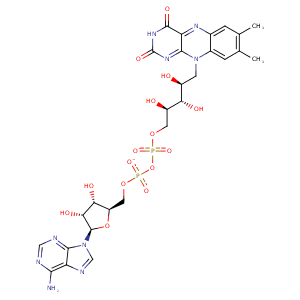
| HET Code: | FAD |
|---|---|
| Formula: | C27H31N9O15P2 |
| Molecular weight: | 783.534 g/mol |
| DrugBank ID: | DB03147 |
| Buried Surface Area: | 73.26 % |
| Polar Surface area: | 381.7 Å2 |
| Number of | |
|---|---|
| H-Bond Acceptors: | 22 |
| H-Bond Donors: | 7 |
| Rings: | 6 |
| Aromatic rings: | 3 |
| Anionic atoms: | 2 |
| Cationic atoms: | 0 |
| Rule of Five Violation: | 3 |
| Rotatable Bonds: | 13 |
| X | Y | Z |
|---|---|---|
| -31.9814 | 91.5819 | 24.0426 |
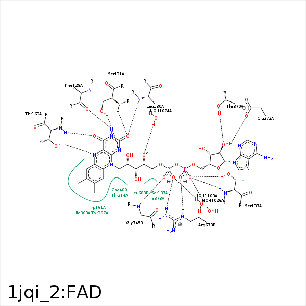


Represent the protein/ligand binding mode, centered on the ligand
Dashed lines represents hydrogen bonds and metal interactions
Green residue labels for amino acids with hydrophobic contacts (green lines) to the ligand
| Ligand | Protein | Interaction | |||
|---|---|---|---|---|---|
| Atom | Atom | Residue | Distance (Å) | Angle (°) | Type |
| N3 | O | PHE- 128 | 2.95 | 154.49 | H-Bond (Ligand Donor) |
| O2 | N | LEU- 130 | 3.02 | 158.65 | H-Bond (Protein Donor) |
| N1 | OG | SER- 131 | 2.99 | 160.37 | H-Bond (Protein Donor) |
| O2 | N | SER- 131 | 3.09 | 175.57 | H-Bond (Protein Donor) |
| C1' | CB | SER- 131 | 3.91 | 0 | Hydrophobic |
| O1A | OG | SER- 137 | 2.72 | 153.38 | H-Bond (Protein Donor) |
| O1A | N | SER- 137 | 3.12 | 153.69 | H-Bond (Protein Donor) |
| C8M | CE3 | TRP- 161 | 4.44 | 0 | Hydrophobic |
| C1' | CB | TRP- 161 | 3.6 | 0 | Hydrophobic |
| C9A | CB | TRP- 161 | 3.26 | 0 | Hydrophobic |
| O4 | OG1 | THR- 163 | 3.22 | 129.06 | H-Bond (Protein Donor) |
| O4 | N | THR- 163 | 2.92 | 167.21 | H-Bond (Protein Donor) |
| N5 | OG1 | THR- 163 | 2.86 | 137.66 | H-Bond (Protein Donor) |
| C7M | CD | LYS- 206 | 3.77 | 0 | Hydrophobic |
| C7M | CD1 | ILE- 209 | 4.04 | 0 | Hydrophobic |
| C6 | CG2 | THR- 214 | 3.92 | 0 | Hydrophobic |
| C8M | CD1 | ILE- 363 | 3.29 | 0 | Hydrophobic |
| C7M | CD1 | ILE- 363 | 3.65 | 0 | Hydrophobic |
| C4' | CG2 | ILE- 366 | 4.46 | 0 | Hydrophobic |
| C7M | CD1 | TYR- 367 | 4.37 | 0 | Hydrophobic |
| C2' | CB | TYR- 367 | 4.34 | 0 | Hydrophobic |
| C9 | CB | TYR- 367 | 4.09 | 0 | Hydrophobic |
| O2B | OG1 | THR- 370 | 2.63 | 161.36 | H-Bond (Protein Donor) |
| C2B | CG2 | THR- 370 | 4.04 | 0 | Hydrophobic |
| C5' | CG2 | THR- 370 | 3.87 | 0 | Hydrophobic |
| O2B | OE1 | GLU- 372 | 2.77 | 132.32 | H-Bond (Ligand Donor) |
| C2B | CD1 | ILE- 373 | 4.43 | 0 | Hydrophobic |
| O2A | CZ | ARG- 673 | 3.53 | 0 | Ionic (Protein Cationic) |
| O2A | NE | ARG- 673 | 2.89 | 154.23 | H-Bond (Protein Donor) |
| O2A | NH2 | ARG- 673 | 3.3 | 134.34 | H-Bond (Protein Donor) |
| O2P | NH2 | ARG- 673 | 2.88 | 124.4 | H-Bond (Protein Donor) |
| C5B | CD1 | LEU- 680 | 3.83 | 0 | Hydrophobic |
| C1B | CD1 | ILE- 686 | 4.22 | 0 | Hydrophobic |
| O1P | N | GLY- 745 | 2.86 | 152.65 | H-Bond (Protein Donor) |
| C7M | CD2 | TYR- 748 | 4.11 | 0 | Hydrophobic |
| C8M | CB | TYR- 748 | 4.08 | 0 | Hydrophobic |
| O1A | O | HOH- 1026 | 2.93 | 179.96 | H-Bond (Protein Donor) |
| O4' | O | HOH- 1074 | 2.91 | 179.96 | H-Bond (Protein Donor) |
| O2P | O | HOH- 1103 | 2.8 | 148.21 | H-Bond (Protein Donor) |
