sc-PDB
An Annotated Database of Druggable Binding Sites from the Protein DataBank
An Annotated Database of Druggable Binding Sites from the Protein DataBank
1.700 Å
X-ray
1995-07-12
| Name: | Trypanothione reductase |
|---|---|
| ID: | TYTR_CRIFA |
| AC: | P39040 |
| Organism: | Crithidia fasciculata |
| Reign: | Eukaryota |
| TaxID: | 5656 |
| EC Number: | 1.8.1.12 |
| Chain Name: | Percentage of Residues within binding site |
|---|---|
| A | 94 % |
| B | 6 % |
| B-Factor: | 15.228 |
|---|---|
| Number of residues: | 82 |
| Including | |
| Standard Amino Acids: | 70 |
| Non Standard Amino Acids: | 0 |
| Water Molecules: | 12 |
| Cofactors: | |
| Metals: | |
| Ligandability | Volume (Å3) |
|---|---|
| 1.269 | 1410.750 |
| % Hydrophobic | % Polar |
|---|---|
| 45.45 | 54.55 |
| According to VolSite | |
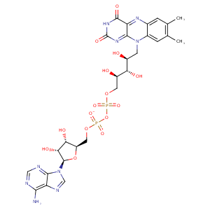
| HET Code: | FAD |
|---|---|
| Formula: | C27H31N9O15P2 |
| Molecular weight: | 783.534 g/mol |
| DrugBank ID: | DB03147 |
| Buried Surface Area: | 80.31 % |
| Polar Surface area: | 381.7 Å2 |
| Number of | |
|---|---|
| H-Bond Acceptors: | 22 |
| H-Bond Donors: | 7 |
| Rings: | 6 |
| Aromatic rings: | 3 |
| Anionic atoms: | 2 |
| Cationic atoms: | 0 |
| Rule of Five Violation: | 3 |
| Rotatable Bonds: | 13 |
| X | Y | Z |
|---|---|---|
| 4.72836 | -53.3741 | -12.8412 |


Represent the protein/ligand binding mode, centered on the ligand
Dashed lines represents hydrogen bonds and metal interactions
Green residue labels for amino acids with hydrophobic contacts (green lines) to the ligand
| Ligand | Protein | Interaction | |||
|---|---|---|---|---|---|
| Atom | Atom | Residue | Distance (Å) | Angle (°) | Type |
| O1A | N | SER- 13 | 3.26 | 166.78 | H-Bond (Protein Donor) |
| C4' | CB | SER- 13 | 4.3 | 0 | Hydrophobic |
| O1P | N | GLY- 14 | 2.83 | 156.43 | H-Bond (Protein Donor) |
| O3B | OD1 | ASP- 34 | 3.35 | 138.24 | H-Bond (Ligand Donor) |
| O3B | OD2 | ASP- 34 | 2.95 | 164.33 | H-Bond (Ligand Donor) |
| O2B | OD1 | ASP- 34 | 2.72 | 152.42 | H-Bond (Ligand Donor) |
| C3B | CB | ALA- 45 | 3.79 | 0 | Hydrophobic |
| O1A | N | THR- 50 | 3.04 | 156.85 | H-Bond (Protein Donor) |
| O2A | OG1 | THR- 50 | 2.69 | 161.06 | H-Bond (Protein Donor) |
| C8M | CG2 | THR- 50 | 3.65 | 0 | Hydrophobic |
| C2' | SG | CYS- 56 | 4.13 | 0 | Hydrophobic |
| N5 | NZ | LYS- 59 | 2.89 | 175.67 | H-Bond (Protein Donor) |
| C6 | CG | LYS- 59 | 4.33 | 0 | Hydrophobic |
| N6A | O | GLY- 126 | 3.2 | 157.53 | H-Bond (Ligand Donor) |
| N1A | N | GLY- 126 | 2.86 | 171.11 | H-Bond (Protein Donor) |
| C7M | CB | SER- 177 | 4 | 0 | Hydrophobic |
| C7M | CE2 | PHE- 181 | 4.09 | 0 | Hydrophobic |
| C6 | CG1 | ILE- 198 | 4.22 | 0 | Hydrophobic |
| C9 | CD1 | ILE- 198 | 4.17 | 0 | Hydrophobic |
| C8 | CD1 | ILE- 198 | 3.65 | 0 | Hydrophobic |
| C7M | CE2 | PHE- 202 | 4.38 | 0 | Hydrophobic |
| C8M | CD | ARG- 286 | 3.97 | 0 | Hydrophobic |
| O3' | OD2 | ASP- 326 | 3.17 | 133.68 | H-Bond (Ligand Donor) |
| O3' | OD1 | ASP- 326 | 2.88 | 165.85 | H-Bond (Ligand Donor) |
| C5' | CB | ASP- 326 | 4.29 | 0 | Hydrophobic |
| O2P | N | ASP- 326 | 2.78 | 150.46 | H-Bond (Protein Donor) |
| O2 | N | THR- 334 | 3 | 150.94 | H-Bond (Protein Donor) |
| C2' | CB | THR- 334 | 4.45 | 0 | Hydrophobic |
| C4' | CB | THR- 334 | 4.41 | 0 | Hydrophobic |
| C5' | CB | ALA- 337 | 4.3 | 0 | Hydrophobic |
| N3 | O | HIS- 460 | 2.83 | 156.66 | H-Bond (Ligand Donor) |
| O1P | O | HOH- 521 | 2.61 | 172.23 | H-Bond (Protein Donor) |
| O2P | O | HOH- 523 | 2.71 | 179.94 | H-Bond (Protein Donor) |
| O2A | O | HOH- 528 | 2.73 | 179.95 | H-Bond (Protein Donor) |
| O2B | O | HOH- 584 | 2.85 | 179.98 | H-Bond (Protein Donor) |
| N6A | O | HOH- 593 | 3.5 | 123.78 | H-Bond (Ligand Donor) |
