sc-PDB
An Annotated Database of Druggable Binding Sites from the Protein DataBank
An Annotated Database of Druggable Binding Sites from the Protein DataBank
1.700 Å
X-ray
1997-03-06
| Name: | Ferredoxin--NADP reductase |
|---|---|
| ID: | FENR_ECOLI |
| AC: | P28861 |
| Organism: | Escherichia coli |
| Reign: | Bacteria |
| TaxID: | 83333 |
| EC Number: | 1.18.1.2 |
| Chain Name: | Percentage of Residues within binding site |
|---|---|
| A | 100 % |
| B-Factor: | 15.584 |
|---|---|
| Number of residues: | 37 |
| Including | |
| Standard Amino Acids: | 34 |
| Non Standard Amino Acids: | 0 |
| Water Molecules: | 3 |
| Cofactors: | |
| Metals: | |
| Ligandability | Volume (Å3) |
|---|---|
| 0.311 | 367.875 |
| % Hydrophobic | % Polar |
|---|---|
| 35.78 | 64.22 |
| According to VolSite | |
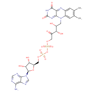
| HET Code: | FAD |
|---|---|
| Formula: | C27H31N9O15P2 |
| Molecular weight: | 783.534 g/mol |
| DrugBank ID: | DB03147 |
| Buried Surface Area: | 57.57 % |
| Polar Surface area: | 381.7 Å2 |
| Number of | |
|---|---|
| H-Bond Acceptors: | 22 |
| H-Bond Donors: | 7 |
| Rings: | 6 |
| Aromatic rings: | 3 |
| Anionic atoms: | 2 |
| Cationic atoms: | 0 |
| Rule of Five Violation: | 3 |
| Rotatable Bonds: | 13 |
| X | Y | Z |
|---|---|---|
| 28.6401 | 24.8455 | 27.975 |
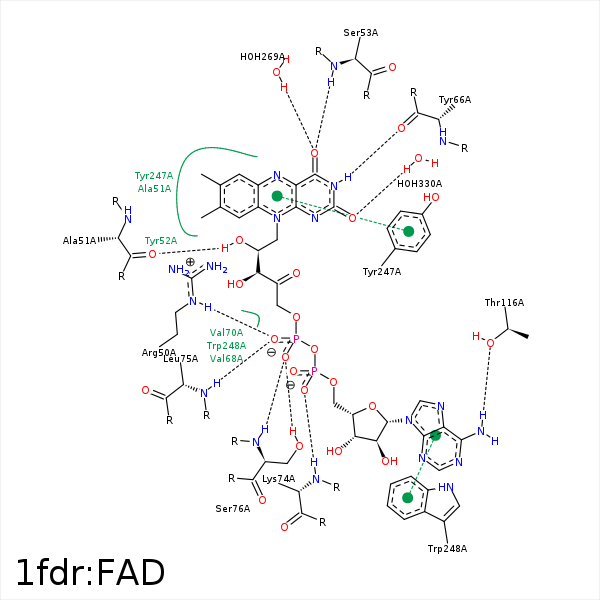


Represent the protein/ligand binding mode, centered on the ligand
Dashed lines represents hydrogen bonds and metal interactions
Green residue labels for amino acids with hydrophobic contacts (green lines) to the ligand
| Ligand | Protein | Interaction | |||
|---|---|---|---|---|---|
| Atom | Atom | Residue | Distance (Å) | Angle (°) | Type |
| C7M | CD2 | PHE- 36 | 3.65 | 0 | Hydrophobic |
| O1A | NH2 | ARG- 50 | 3.16 | 131.04 | H-Bond (Protein Donor) |
| O1A | NE | ARG- 50 | 3.19 | 125.8 | H-Bond (Protein Donor) |
| O1P | NH2 | ARG- 50 | 3.38 | 121.05 | H-Bond (Protein Donor) |
| O1P | NE | ARG- 50 | 2.89 | 139.68 | H-Bond (Protein Donor) |
| O1A | CZ | ARG- 50 | 3.56 | 0 | Ionic (Protein Cationic) |
| O1P | CZ | ARG- 50 | 3.5 | 0 | Ionic (Protein Cationic) |
| C3' | CB | ARG- 50 | 4.09 | 0 | Hydrophobic |
| O2' | O | ALA- 51 | 2.7 | 162.47 | H-Bond (Ligand Donor) |
| C7 | CB | ALA- 51 | 3.82 | 0 | Hydrophobic |
| C8 | CB | ALA- 51 | 3.86 | 0 | Hydrophobic |
| C2' | CE1 | TYR- 52 | 4.01 | 0 | Hydrophobic |
| O4 | N | SER- 53 | 3.22 | 139.22 | H-Bond (Protein Donor) |
| N5 | N | SER- 53 | 3.36 | 142.35 | H-Bond (Protein Donor) |
| N3 | O | TYR- 66 | 2.73 | 175.09 | H-Bond (Ligand Donor) |
| O2 | N | VAL- 68 | 3.48 | 134.69 | H-Bond (Protein Donor) |
| C4B | CG1 | VAL- 70 | 4.24 | 0 | Hydrophobic |
| C1B | CG1 | VAL- 70 | 3.97 | 0 | Hydrophobic |
| C5' | CG2 | VAL- 70 | 3.76 | 0 | Hydrophobic |
| O2A | N | LYS- 74 | 3.21 | 146.97 | H-Bond (Protein Donor) |
| O1P | N | LEU- 75 | 2.93 | 164.65 | H-Bond (Protein Donor) |
| C5' | CB | SER- 76 | 4.25 | 0 | Hydrophobic |
| O2P | N | SER- 76 | 2.91 | 149.18 | H-Bond (Protein Donor) |
| N6A | OG1 | THR- 116 | 3.1 | 142.93 | H-Bond (Ligand Donor) |
| C7M | CG | GLU- 245 | 4.31 | 0 | Hydrophobic |
| C1' | CB | TYR- 247 | 4.22 | 0 | Hydrophobic |
| C9A | CB | TYR- 247 | 4 | 0 | Hydrophobic |
| C3B | CB | TRP- 248 | 3.87 | 0 | Hydrophobic |
| C2B | CD2 | TRP- 248 | 3.74 | 0 | Hydrophobic |
| O4 | O | HOH- 269 | 2.52 | 149.28 | H-Bond (Protein Donor) |
| O2 | O | HOH- 330 | 2.88 | 138.3 | H-Bond (Protein Donor) |
