sc-PDB
An Annotated Database of Druggable Binding Sites from the Protein DataBank
An Annotated Database of Druggable Binding Sites from the Protein DataBank
1.850 Å
X-ray
2000-03-02
| Name: | 6,7-dimethyl-8-ribityllumazine synthase |
|---|---|
| ID: | RIB4_YEAST |
| AC: | P50861 |
| Organism: | Saccharomyces cerevisiae |
| Reign: | Eukaryota |
| TaxID: | 559292 |
| EC Number: | 2.5.1.78 |
| Chain Name: | Percentage of Residues within binding site |
|---|---|
| C | 67 % |
| D | 33 % |
| B-Factor: | 19.987 |
|---|---|
| Number of residues: | 38 |
| Including | |
| Standard Amino Acids: | 36 |
| Non Standard Amino Acids: | 0 |
| Water Molecules: | 2 |
| Cofactors: | |
| Metals: | |
| Ligandability | Volume (Å3) |
|---|---|
| 0.816 | 810.000 |
| % Hydrophobic | % Polar |
|---|---|
| 55.83 | 44.17 |
| According to VolSite | |
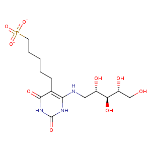
| HET Code: | INJ |
|---|---|
| Formula: | C14H24N3O9P |
| Molecular weight: | 409.329 g/mol |
| DrugBank ID: | DB04266 |
| Buried Surface Area: | 70.53 % |
| Polar Surface area: | 232.19 Å2 |
| Number of | |
|---|---|
| H-Bond Acceptors: | 12 |
| H-Bond Donors: | 7 |
| Rings: | 1 |
| Aromatic rings: | 1 |
| Anionic atoms: | 2 |
| Cationic atoms: | 0 |
| Rule of Five Violation: | 2 |
| Rotatable Bonds: | 12 |
| X | Y | Z |
|---|---|---|
| 67.1905 | 32.4042 | 41.3084 |
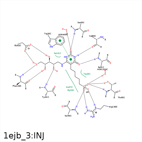


Represent the protein/ligand binding mode, centered on the ligand
Dashed lines represents hydrogen bonds and metal interactions
Green residue labels for amino acids with hydrophobic contacts (green lines) to the ligand
| Ligand | Protein | Interaction | |||
|---|---|---|---|---|---|
| Atom | Atom | Residue | Distance (Å) | Angle (°) | Type |
| C8 | CE2 | TRP- 26 | 3.55 | 0 | Hydrophobic |
| C13 | CZ3 | TRP- 26 | 3.52 | 0 | Hydrophobic |
| O2 | N | SER- 60 | 2.82 | 143.69 | H-Bond (Protein Donor) |
| O9 | N | TYR- 61 | 3.05 | 170.73 | H-Bond (Protein Donor) |
| C10 | CB | TYR- 61 | 3.81 | 0 | Hydrophobic |
| O10 | OE1 | GLU- 62 | 2.55 | 172.66 | H-Bond (Ligand Donor) |
| O12 | OE2 | GLU- 62 | 2.61 | 160.8 | H-Bond (Ligand Donor) |
| N3 | O | VAL- 89 | 2.92 | 177.27 | H-Bond (Ligand Donor) |
| C14 | CD2 | LEU- 90 | 4.24 | 0 | Hydrophobic |
| O4 | N | ILE- 91 | 3.27 | 160.35 | H-Bond (Protein Donor) |
| O3P | N | SER- 94 | 2.89 | 165.35 | H-Bond (Protein Donor) |
| O2P | OG1 | THR- 95 | 2.53 | 171.49 | H-Bond (Protein Donor) |
| O2P | N | THR- 95 | 2.79 | 151.37 | H-Bond (Protein Donor) |
| C14 | CB | HIS- 97 | 4.45 | 0 | Hydrophobic |
| C16 | CB | HIS- 97 | 4.03 | 0 | Hydrophobic |
| C15 | CB | PHE- 98 | 4.43 | 0 | Hydrophobic |
| C14 | CD1 | ILE- 101 | 4.19 | 0 | Hydrophobic |
| C9 | CD1 | ILE- 101 | 3.93 | 0 | Hydrophobic |
| C12 | CG2 | ILE- 121 | 3.48 | 0 | Hydrophobic |
| O11 | O | PHE- 122 | 3.02 | 166.49 | H-Bond (Ligand Donor) |
| C12 | CB | PHE- 122 | 4.16 | 0 | Hydrophobic |
| O12 | N | PHE- 122 | 2.8 | 150.49 | H-Bond (Protein Donor) |
| O1P | CZ | ARG- 136 | 3.44 | 0 | Ionic (Protein Cationic) |
| O3P | CZ | ARG- 136 | 3.69 | 0 | Ionic (Protein Cationic) |
| O1P | NE | ARG- 136 | 2.66 | 174.72 | H-Bond (Protein Donor) |
| O1P | NH2 | ARG- 136 | 3.35 | 129.34 | H-Bond (Protein Donor) |
| O3P | NH2 | ARG- 136 | 2.73 | 168.21 | H-Bond (Protein Donor) |
| O2 | O | HOH- 409 | 3.03 | 179.94 | H-Bond (Protein Donor) |
| O2P | O | HOH- 1310 | 2.74 | 179.96 | H-Bond (Protein Donor) |
