sc-PDB
An Annotated Database of Druggable Binding Sites from the Protein DataBank
An Annotated Database of Druggable Binding Sites from the Protein DataBank
1.750 Å
X-ray
2000-02-11
| Name: | Modification methylase RsrI |
|---|---|
| ID: | MTR1_RHOSH |
| AC: | P14751 |
| Organism: | Rhodobacter sphaeroides |
| Reign: | Bacteria |
| TaxID: | 1063 |
| EC Number: | 2.1.1.72 |
| Chain Name: | Percentage of Residues within binding site |
|---|---|
| A | 100 % |
| B-Factor: | 19.754 |
|---|---|
| Number of residues: | 27 |
| Including | |
| Standard Amino Acids: | 25 |
| Non Standard Amino Acids: | 0 |
| Water Molecules: | 2 |
| Cofactors: | |
| Metals: | |
| Ligandability | Volume (Å3) |
|---|---|
| 0.097 | 394.875 |
| % Hydrophobic | % Polar |
|---|---|
| 41.88 | 58.12 |
| According to VolSite | |
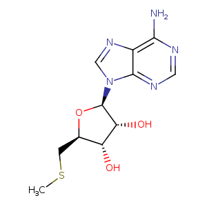
| HET Code: | MTA |
|---|---|
| Formula: | C11H15N5O3S |
| Molecular weight: | 297.333 g/mol |
| DrugBank ID: | DB02282 |
| Buried Surface Area: | 60.18 % |
| Polar Surface area: | 144.61 Å2 |
| Number of | |
|---|---|
| H-Bond Acceptors: | 8 |
| H-Bond Donors: | 3 |
| Rings: | 3 |
| Aromatic rings: | 2 |
| Anionic atoms: | 0 |
| Cationic atoms: | 0 |
| Rule of Five Violation: | 0 |
| Rotatable Bonds: | 3 |
| X | Y | Z |
|---|---|---|
| 27.4844 | 24.9794 | -4.206 |
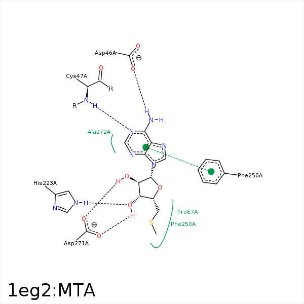


Represent the protein/ligand binding mode, centered on the ligand
Dashed lines represents hydrogen bonds and metal interactions
Green residue labels for amino acids with hydrophobic contacts (green lines) to the ligand
| Ligand | Protein | Interaction | |||
|---|---|---|---|---|---|
| Atom | Atom | Residue | Distance (Å) | Angle (°) | Type |
| N6 | OD2 | ASP- 46 | 2.75 | 139.56 | H-Bond (Ligand Donor) |
| N1 | N | CYS- 47 | 2.88 | 153.35 | H-Bond (Protein Donor) |
| CS | CB | ASP- 65 | 4.09 | 0 | Hydrophobic |
| O3' | NE2 | HIS- 223 | 3.06 | 160.88 | H-Bond (Protein Donor) |
| C1' | CD1 | PHE- 250 | 4.12 | 0 | Hydrophobic |
| C4' | CB | PHE- 250 | 3.97 | 0 | Hydrophobic |
| O2' | OD1 | ASP- 271 | 2.69 | 163.6 | H-Bond (Ligand Donor) |
| O3' | OD2 | ASP- 271 | 2.74 | 141.71 | H-Bond (Ligand Donor) |
| N3 | N | ALA- 272 | 3.29 | 145.3 | H-Bond (Protein Donor) |
