sc-PDB
An Annotated Database of Druggable Binding Sites from the Protein DataBank
An Annotated Database of Druggable Binding Sites from the Protein DataBank
2.400 Å
X-ray
1992-03-14
| Name: | Dihydrofolate reductase |
|---|---|
| ID: | DYR_CHICK |
| AC: | P00378 |
| Organism: | Gallus gallus |
| Reign: | Eukaryota |
| TaxID: | 9031 |
| EC Number: | 1.5.1.3 |
| Chain Name: | Percentage of Residues within binding site |
|---|---|
| A | 100 % |
| B-Factor: | 24.847 |
|---|---|
| Number of residues: | 27 |
| Including | |
| Standard Amino Acids: | 26 |
| Non Standard Amino Acids: | 1 |
| Water Molecules: | 0 |
| Cofactors: | NAP |
| Metals: | |
| Ligandability | Volume (Å3) |
|---|---|
| 1.244 | 455.625 |
| % Hydrophobic | % Polar |
|---|---|
| 62.22 | 37.78 |
| According to VolSite | |
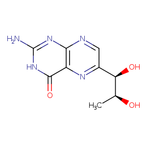
| HET Code: | HBI |
|---|---|
| Formula: | C9H13N5O3 |
| Molecular weight: | 239.231 g/mol |
| DrugBank ID: | DB04400 |
| Buried Surface Area: | 65.47 % |
| Polar Surface area: | 132.33 Å2 |
| Number of | |
|---|---|
| H-Bond Acceptors: | 7 |
| H-Bond Donors: | 5 |
| Rings: | 2 |
| Aromatic rings: | 0 |
| Anionic atoms: | 0 |
| Cationic atoms: | 0 |
| Rule of Five Violation: | 0 |
| Rotatable Bonds: | 2 |
| X | Y | Z |
|---|---|---|
| 17.3186 | 7.36735 | 7.183 |
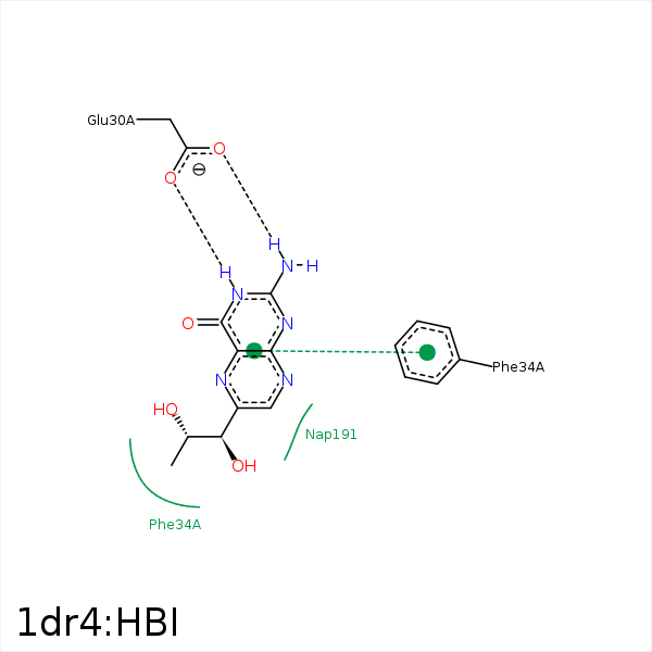


Represent the protein/ligand binding mode, centered on the ligand
Dashed lines represents hydrogen bonds and metal interactions
Green residue labels for amino acids with hydrophobic contacts (green lines) to the ligand
| Ligand | Protein | Interaction | |||
|---|---|---|---|---|---|
| Atom | Atom | Residue | Distance (Å) | Angle (°) | Type |
| N2 | OE2 | GLU- 30 | 2.76 | 175.05 | H-Bond (Ligand Donor) |
| N3 | OE1 | GLU- 30 | 2.84 | 179.73 | H-Bond (Ligand Donor) |
| C11 | CZ | PHE- 34 | 3.63 | 0 | Hydrophobic |
| C9 | CG2 | THR- 56 | 4.07 | 0 | Hydrophobic |
| C11 | CD1 | LEU- 67 | 4.22 | 0 | Hydrophobic |
| C11 | CG2 | VAL- 115 | 4.41 | 0 | Hydrophobic |
| C9 | C4N | NAP- 191 | 3.64 | 0 | Hydrophobic |
