sc-PDB
An Annotated Database of Druggable Binding Sites from the Protein DataBank
An Annotated Database of Druggable Binding Sites from the Protein DataBank
1.900 Å
X-ray
1999-03-23
| Name: | Glucose oxidase |
|---|---|
| ID: | GOX_ASPNG |
| AC: | P13006 |
| Organism: | Aspergillus niger |
| Reign: | Eukaryota |
| TaxID: | 5061 |
| EC Number: | 1.1.3.4 |
| Chain Name: | Percentage of Residues within binding site |
|---|---|
| A | 100 % |
| B-Factor: | 16.488 |
|---|---|
| Number of residues: | 66 |
| Including | |
| Standard Amino Acids: | 63 |
| Non Standard Amino Acids: | 0 |
| Water Molecules: | 3 |
| Cofactors: | |
| Metals: | |
| Ligandability | Volume (Å3) |
|---|---|
| 1.086 | 1400.625 |
| % Hydrophobic | % Polar |
|---|---|
| 42.17 | 57.83 |
| According to VolSite | |
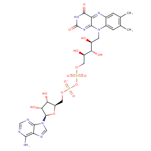
| HET Code: | FAD |
|---|---|
| Formula: | C27H31N9O15P2 |
| Molecular weight: | 783.534 g/mol |
| DrugBank ID: | DB03147 |
| Buried Surface Area: | 79.4 % |
| Polar Surface area: | 381.7 Å2 |
| Number of | |
|---|---|
| H-Bond Acceptors: | 22 |
| H-Bond Donors: | 7 |
| Rings: | 6 |
| Aromatic rings: | 3 |
| Anionic atoms: | 2 |
| Cationic atoms: | 0 |
| Rule of Five Violation: | 3 |
| Rotatable Bonds: | 13 |
| X | Y | Z |
|---|---|---|
| 33.178 | 7.16268 | 57.7585 |
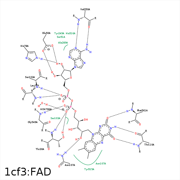


Represent the protein/ligand binding mode, centered on the ligand
Dashed lines represents hydrogen bonds and metal interactions
Green residue labels for amino acids with hydrophobic contacts (green lines) to the ligand
| Ligand | Protein | Interaction | |||
|---|---|---|---|---|---|
| Atom | Atom | Residue | Distance (Å) | Angle (°) | Type |
| O2A | N | LEU- 29 | 3.24 | 173.33 | H-Bond (Protein Donor) |
| C4' | CD1 | LEU- 29 | 3.95 | 0 | Hydrophobic |
| O1P | OG1 | THR- 30 | 2.85 | 169.98 | H-Bond (Protein Donor) |
| O1P | N | THR- 30 | 2.81 | 166.89 | H-Bond (Protein Donor) |
| O3B | OE1 | GLU- 50 | 2.79 | 169.22 | H-Bond (Ligand Donor) |
| O2B | OE2 | GLU- 50 | 2.66 | 160.55 | H-Bond (Ligand Donor) |
| N3A | N | SER- 51 | 3.46 | 136.36 | H-Bond (Protein Donor) |
| C7M | CE1 | TYR- 68 | 4.21 | 0 | Hydrophobic |
| C7M | CZ | PHE- 72 | 3.88 | 0 | Hydrophobic |
| O2B | NE2 | HIS- 78 | 2.81 | 177.39 | H-Bond (Protein Donor) |
| C7M | CB | ARG- 95 | 4.27 | 0 | Hydrophobic |
| C8M | CB | ARG- 95 | 4.5 | 0 | Hydrophobic |
| O1A | OG | SER- 103 | 3.42 | 164.8 | H-Bond (Protein Donor) |
| O2A | N | SER- 103 | 3.19 | 151.32 | H-Bond (Protein Donor) |
| C3' | CB | SER- 103 | 4.18 | 0 | Hydrophobic |
| C8M | CB | SER- 103 | 4.08 | 0 | Hydrophobic |
| C7M | CG2 | VAL- 106 | 4.4 | 0 | Hydrophobic |
| C8M | CG2 | VAL- 106 | 4.44 | 0 | Hydrophobic |
| O2' | ND2 | ASN- 107 | 3.04 | 160.38 | H-Bond (Protein Donor) |
| C9A | CB | ASN- 107 | 3.48 | 0 | Hydrophobic |
| N3 | O | THR- 110 | 3 | 156.96 | H-Bond (Ligand Donor) |
| O4 | N | THR- 110 | 3.17 | 150.58 | H-Bond (Protein Donor) |
| N6A | O | VAL- 250 | 3.15 | 153.93 | H-Bond (Ligand Donor) |
| N1A | N | VAL- 250 | 2.94 | 162.59 | H-Bond (Protein Donor) |
| C7M | CD2 | TYR- 515 | 3.49 | 0 | Hydrophobic |
| C8M | CB | TYR- 515 | 3.58 | 0 | Hydrophobic |
| O2P | N | GLY- 549 | 3.09 | 166.73 | H-Bond (Protein Donor) |
| C1' | CG2 | VAL- 560 | 4.03 | 0 | Hydrophobic |
| O2 | N | MET- 561 | 2.93 | 161.23 | H-Bond (Protein Donor) |
| C2' | CG | MET- 561 | 4.31 | 0 | Hydrophobic |
| C5' | CD2 | PHE- 564 | 3.83 | 0 | Hydrophobic |
| O1P | O | HOH- 786 | 2.88 | 179.97 | H-Bond (Protein Donor) |
| O2 | O | HOH- 902 | 3.26 | 138.01 | H-Bond (Protein Donor) |
