sc-PDB
An Annotated Database of Druggable Binding Sites from the Protein DataBank
An Annotated Database of Druggable Binding Sites from the Protein DataBank
2.200 Å
X-ray
1998-11-25
| Name: | Alcohol dehydrogenase |
|---|---|
| ID: | ADH_DROLE |
| AC: | P10807 |
| Organism: | Drosophila lebanonensis |
| Reign: | Eukaryota |
| TaxID: | 7225 |
| EC Number: | 1.1.1.1 |
| Chain Name: | Percentage of Residues within binding site |
|---|---|
| A | 2 % |
| B | 98 % |
| B-Factor: | 22.643 |
|---|---|
| Number of residues: | 49 |
| Including | |
| Standard Amino Acids: | 48 |
| Non Standard Amino Acids: | 0 |
| Water Molecules: | 1 |
| Cofactors: | |
| Metals: | |
| Ligandability | Volume (Å3) |
|---|---|
| 1.111 | 678.375 |
| % Hydrophobic | % Polar |
|---|---|
| 50.25 | 49.75 |
| According to VolSite | |
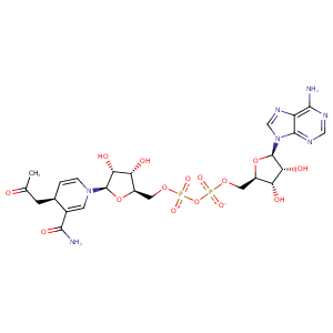
| HET Code: | NAE |
|---|---|
| Formula: | C24H30N7O15P2 |
| Molecular weight: | 718.480 g/mol |
| DrugBank ID: | DB02732 |
| Buried Surface Area: | 72.98 % |
| Polar Surface area: | 360.61 Å2 |
| Number of | |
|---|---|
| H-Bond Acceptors: | 19 |
| H-Bond Donors: | 6 |
| Rings: | 5 |
| Aromatic rings: | 3 |
| Anionic atoms: | 2 |
| Cationic atoms: | 1 |
| Rule of Five Violation: | 3 |
| Rotatable Bonds: | 13 |
| X | Y | Z |
|---|---|---|
| 15.956 | 3.17462 | 30.909 |
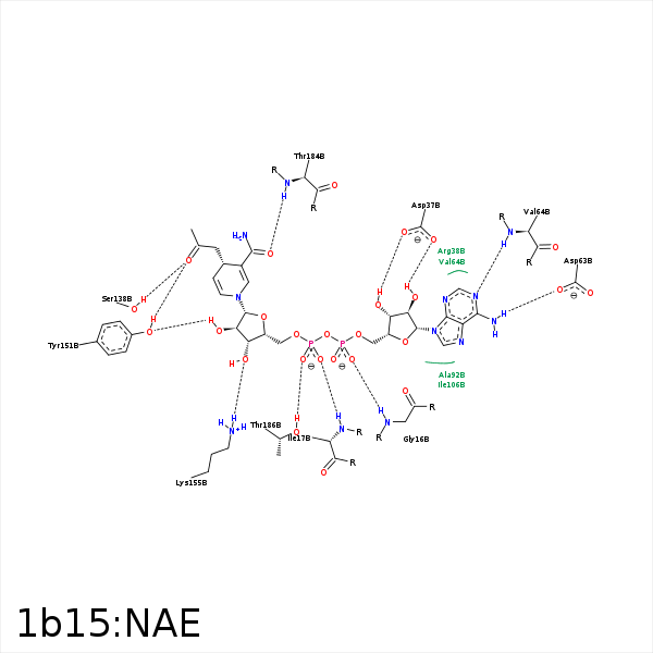


Represent the protein/ligand binding mode, centered on the ligand
Dashed lines represents hydrogen bonds and metal interactions
Green residue labels for amino acids with hydrophobic contacts (green lines) to the ligand
| Ligand | Protein | Interaction | |||
|---|---|---|---|---|---|
| Atom | Atom | Residue | Distance (Å) | Angle (°) | Type |
| C1' | CB | ALA- 12 | 3.45 | 0 | Hydrophobic |
| C4' | CB | ALA- 12 | 3.93 | 0 | Hydrophobic |
| O2A | N | GLY- 16 | 2.88 | 171.37 | H-Bond (Protein Donor) |
| O2N | N | ILE- 17 | 2.83 | 164.29 | H-Bond (Protein Donor) |
| C5B | CB | ILE- 17 | 4.34 | 0 | Hydrophobic |
| O3' | OD1 | ASP- 37 | 3.32 | 143.29 | H-Bond (Ligand Donor) |
| O3' | OD2 | ASP- 37 | 2.68 | 132.49 | H-Bond (Ligand Donor) |
| N6A | OD1 | ASP- 63 | 2.83 | 158.22 | H-Bond (Ligand Donor) |
| N1A | N | VAL- 64 | 3.07 | 165.51 | H-Bond (Protein Donor) |
| C1' | CB | ALA- 92 | 4.31 | 0 | Hydrophobic |
| O4' | N | GLY- 93 | 3.47 | 133.48 | H-Bond (Protein Donor) |
| C2P | CD2 | LEU- 95 | 4.44 | 0 | Hydrophobic |
| C4B | CG2 | ILE- 136 | 3.82 | 0 | Hydrophobic |
| C1B | CG2 | ILE- 136 | 4.13 | 0 | Hydrophobic |
| OP | OG | SER- 138 | 2.78 | 144.76 | H-Bond (Protein Donor) |
| O2B | OH | TYR- 151 | 2.87 | 163.01 | H-Bond (Ligand Donor) |
| C2P | CE2 | TYR- 151 | 4.05 | 0 | Hydrophobic |
| O3B | NZ | LYS- 155 | 3.09 | 145.16 | H-Bond (Protein Donor) |
| O2B | NZ | LYS- 155 | 3.2 | 127.91 | H-Bond (Protein Donor) |
| C4N | CB | PRO- 181 | 3.83 | 0 | Hydrophobic |
| C4P | CD1 | ILE- 183 | 3.97 | 0 | Hydrophobic |
| C2P | CD1 | ILE- 183 | 4.16 | 0 | Hydrophobic |
| O7N | N | THR- 184 | 2.58 | 144.28 | H-Bond (Protein Donor) |
| N7N | O | THR- 184 | 3.21 | 133.42 | H-Bond (Ligand Donor) |
| O1N | OG1 | THR- 186 | 2.68 | 158.05 | H-Bond (Protein Donor) |
| C2B | CD2 | LEU- 188 | 4.06 | 0 | Hydrophobic |
| C2P | CD2 | LEU- 188 | 4.08 | 0 | Hydrophobic |
| C2P | CG1 | VAL- 189 | 3.82 | 0 | Hydrophobic |
| O3B | O | HOH- 266 | 3.49 | 136.01 | H-Bond (Protein Donor) |
