sc-PDB
An Annotated Database of Druggable Binding Sites from the Protein DataBank
An Annotated Database of Druggable Binding Sites from the Protein DataBank
2.000 Å
X-ray
1997-06-20
| Name: | ATP-dependent molecular chaperone HSP82 |
|---|---|
| ID: | HSP82_YEAST |
| AC: | P02829 |
| Organism: | Saccharomyces cerevisiae |
| Reign: | Eukaryota |
| TaxID: | 559292 |
| EC Number: | / |
| Chain Name: | Percentage of Residues within binding site |
|---|---|
| A | 100 % |
| B-Factor: | 16.401 |
|---|---|
| Number of residues: | 36 |
| Including | |
| Standard Amino Acids: | 34 |
| Non Standard Amino Acids: | 0 |
| Water Molecules: | 2 |
| Cofactors: | |
| Metals: | |
| Ligandability | Volume (Å3) |
|---|---|
| 1.046 | 540.000 |
| % Hydrophobic | % Polar |
|---|---|
| 45.63 | 54.38 |
| According to VolSite | |
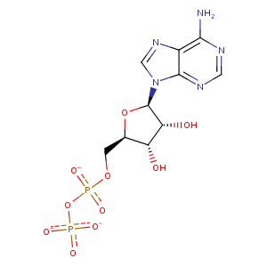
| HET Code: | ADP |
|---|---|
| Formula: | C10H12N5O10P2 |
| Molecular weight: | 424.177 g/mol |
| DrugBank ID: | - |
| Buried Surface Area: | 55.12 % |
| Polar Surface area: | 260.7 Å2 |
| Number of | |
|---|---|
| H-Bond Acceptors: | 14 |
| H-Bond Donors: | 3 |
| Rings: | 3 |
| Aromatic rings: | 2 |
| Anionic atoms: | 3 |
| Cationic atoms: | 0 |
| Rule of Five Violation: | 1 |
| Rotatable Bonds: | 6 |
| X | Y | Z |
|---|---|---|
| 14.4728 | -3.0783 | -3.839 |
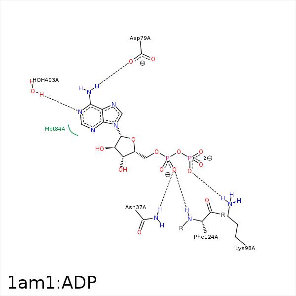


Represent the protein/ligand binding mode, centered on the ligand
Dashed lines represents hydrogen bonds and metal interactions
Green residue labels for amino acids with hydrophobic contacts (green lines) to the ligand
| Ligand | Protein | Interaction | |||
|---|---|---|---|---|---|
| Atom | Atom | Residue | Distance (Å) | Angle (°) | Type |
| O2A | ND2 | ASN- 37 | 2.8 | 147.47 | H-Bond (Protein Donor) |
| N6 | OD2 | ASP- 79 | 2.93 | 162.54 | H-Bond (Ligand Donor) |
| C1' | SD | MET- 84 | 3.88 | 0 | Hydrophobic |
| C4' | CB | ASN- 92 | 4.06 | 0 | Hydrophobic |
| C1' | CB | ASN- 92 | 4.12 | 0 | Hydrophobic |
| C1' | CD2 | LEU- 93 | 4.42 | 0 | Hydrophobic |
| C5' | CD2 | LEU- 93 | 4.11 | 0 | Hydrophobic |
| O1B | NZ | LYS- 98 | 3.2 | 148.4 | H-Bond (Protein Donor) |
| O1B | NZ | LYS- 98 | 3.2 | 0 | Ionic (Protein Cationic) |
| O2A | N | PHE- 124 | 3 | 131.11 | H-Bond (Protein Donor) |
| C5' | CB | PHE- 124 | 4.38 | 0 | Hydrophobic |
| N1 | O | HOH- 403 | 2.76 | 165.67 | H-Bond (Protein Donor) |
