sc-PDB
An Annotated Database of Druggable Binding Sites from the Protein DataBank
An Annotated Database of Druggable Binding Sites from the Protein DataBank
2.100 Å
X-ray
2016-06-18
| Name: | L-threonine 3-dehydrogenase |
|---|---|
| ID: | Q7YW97_9TRYP |
| AC: | Q7YW97 |
| Organism: | Trypanosoma brucei |
| Reign: | Eukaryota |
| TaxID: | 5691 |
| EC Number: | / |
| Chain Name: | Percentage of Residues within binding site |
|---|---|
| D | 100 % |
| B-Factor: | 14.797 |
|---|---|
| Number of residues: | 51 |
| Including | |
| Standard Amino Acids: | 49 |
| Non Standard Amino Acids: | 0 |
| Water Molecules: | 2 |
| Cofactors: | |
| Metals: | |
| Ligandability | Volume (Å3) |
|---|---|
| 1.339 | 796.500 |
| % Hydrophobic | % Polar |
|---|---|
| 51.27 | 48.73 |
| According to VolSite | |
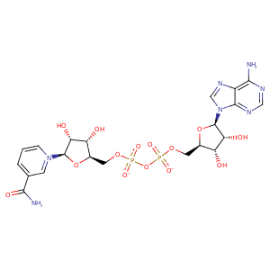
| HET Code: | NAD |
|---|---|
| Formula: | C21H26N7O14P2 |
| Molecular weight: | 662.417 g/mol |
| DrugBank ID: | - |
| Buried Surface Area: | 71.06 % |
| Polar Surface area: | 343.54 Å2 |
| Number of | |
|---|---|
| H-Bond Acceptors: | 18 |
| H-Bond Donors: | 6 |
| Rings: | 5 |
| Aromatic rings: | 3 |
| Anionic atoms: | 2 |
| Cationic atoms: | 1 |
| Rule of Five Violation: | 3 |
| Rotatable Bonds: | 11 |
| X | Y | Z |
|---|---|---|
| 90.0755 | 187.372 | 12.1622 |
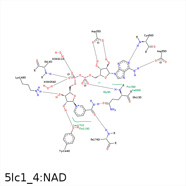


Represent the protein/ligand binding mode, centered on the ligand
Dashed lines represents hydrogen bonds and metal interactions
Green residue labels for amino acids with hydrophobic contacts (green lines) to the ligand
| Ligand | Protein | Interaction | |||
|---|---|---|---|---|---|
| Atom | Atom | Residue | Distance (Å) | Angle (°) | Type |
| O1A | N | GLN- 13 | 2.81 | 170.88 | H-Bond (Protein Donor) |
| N7N | OE1 | GLN- 13 | 3.12 | 154.23 | H-Bond (Ligand Donor) |
| O1N | N | ILE- 14 | 2.86 | 170.63 | H-Bond (Protein Donor) |
| C5D | CG1 | ILE- 14 | 3.94 | 0 | Hydrophobic |
| C3N | CD1 | ILE- 14 | 4.5 | 0 | Hydrophobic |
| O3B | OD1 | ASP- 35 | 3.4 | 126.18 | H-Bond (Ligand Donor) |
| O2B | OD1 | ASP- 35 | 2.52 | 158.98 | H-Bond (Ligand Donor) |
| C1B | CG1 | VAL- 36 | 4.48 | 0 | Hydrophobic |
| N3A | N | VAL- 36 | 3.46 | 140.63 | H-Bond (Protein Donor) |
| N6A | OD1 | ASP- 55 | 2.93 | 138.78 | H-Bond (Ligand Donor) |
| N1A | N | CYS- 56 | 2.96 | 168.81 | H-Bond (Protein Donor) |
| O3D | O | LEU- 77 | 3.22 | 123.86 | H-Bond (Ligand Donor) |
| C5D | CB | LEU- 77 | 3.96 | 0 | Hydrophobic |
| C1B | CB | PRO- 78 | 4.42 | 0 | Hydrophobic |
| C3D | CB | ALA- 79 | 3.61 | 0 | Hydrophobic |
| C2D | SD | MET- 81 | 3.94 | 0 | Hydrophobic |
| C3N | SD | MET- 81 | 4.28 | 0 | Hydrophobic |
| C4D | CG | PRO- 117 | 3.61 | 0 | Hydrophobic |
| C5N | CB | THR- 119 | 3.64 | 0 | Hydrophobic |
| O2D | OH | TYR- 144 | 2.55 | 148.79 | H-Bond (Ligand Donor) |
| O3D | NZ | LYS- 148 | 2.97 | 166.42 | H-Bond (Protein Donor) |
| C5N | CB | LEU- 171 | 3.44 | 0 | Hydrophobic |
| O7N | N | ILE- 174 | 2.95 | 159.5 | H-Bond (Protein Donor) |
| C3N | CG1 | ILE- 174 | 4.29 | 0 | Hydrophobic |
| O2N | O | HOH- 611 | 2.55 | 179.96 | H-Bond (Protein Donor) |
| O1N | O | HOH- 654 | 2.68 | 122.93 | H-Bond (Protein Donor) |
