sc-PDB
An Annotated Database of Druggable Binding Sites from the Protein DataBank
An Annotated Database of Druggable Binding Sites from the Protein DataBank
2.300 Å
X-ray
2016-01-10
| Name: | UDP-galactopyranose mutase |
|---|---|
| ID: | Q4W1X2_ASPFM |
| AC: | Q4W1X2 |
| Organism: | Neosartorya fumigata |
| Reign: | Eukaryota |
| TaxID: | 746128 |
| EC Number: | / |
| Chain Name: | Percentage of Residues within binding site |
|---|---|
| D | 100 % |
| B-Factor: | 43.182 |
|---|---|
| Number of residues: | 61 |
| Including | |
| Standard Amino Acids: | 59 |
| Non Standard Amino Acids: | 0 |
| Water Molecules: | 2 |
| Cofactors: | |
| Metals: | |
| Ligandability | Volume (Å3) |
|---|---|
| 1.005 | 1505.250 |
| % Hydrophobic | % Polar |
|---|---|
| 37.44 | 62.56 |
| According to VolSite | |
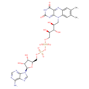
| HET Code: | FDA |
|---|---|
| Formula: | C27H33N9O15P2 |
| Molecular weight: | 785.550 g/mol |
| DrugBank ID: | - |
| Buried Surface Area: | 65.17 % |
| Polar Surface area: | 381.04 Å2 |
| Number of | |
|---|---|
| H-Bond Acceptors: | 21 |
| H-Bond Donors: | 9 |
| Rings: | 6 |
| Aromatic rings: | 3 |
| Anionic atoms: | 2 |
| Cationic atoms: | 0 |
| Rule of Five Violation: | 3 |
| Rotatable Bonds: | 13 |
| X | Y | Z |
|---|---|---|
| 36.0719 | 58.2929 | 228.317 |
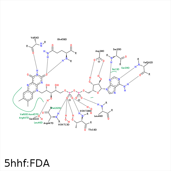


Represent the protein/ligand binding mode, centered on the ligand
Dashed lines represents hydrogen bonds and metal interactions
Green residue labels for amino acids with hydrophobic contacts (green lines) to the ligand
| Ligand | Protein | Interaction | |||
|---|---|---|---|---|---|
| Atom | Atom | Residue | Distance (Å) | Angle (°) | Type |
| C4' | CG | PRO- 17 | 3.91 | 0 | Hydrophobic |
| O1P | OG1 | THR- 18 | 2.88 | 162.73 | H-Bond (Protein Donor) |
| O2P | OG1 | THR- 18 | 3.45 | 121.5 | H-Bond (Protein Donor) |
| O2P | N | THR- 18 | 3.04 | 161.34 | H-Bond (Protein Donor) |
| O3B | OD1 | ASP- 38 | 3.49 | 131.06 | H-Bond (Ligand Donor) |
| O3B | OD2 | ASP- 38 | 2.58 | 164.42 | H-Bond (Ligand Donor) |
| O2B | OD1 | ASP- 38 | 3.26 | 164.45 | H-Bond (Ligand Donor) |
| N3A | N | SER- 39 | 3.12 | 141.13 | H-Bond (Protein Donor) |
| O2A | N | LEU- 46 | 2.84 | 158.34 | H-Bond (Protein Donor) |
| C8M | CD1 | LEU- 46 | 3.59 | 0 | Hydrophobic |
| C4' | CB | LEU- 46 | 4.49 | 0 | Hydrophobic |
| C8M | CG1 | VAL- 60 | 3.74 | 0 | Hydrophobic |
| N3 | O | VAL- 64 | 3.09 | 157.8 | H-Bond (Ligand Donor) |
| N6A | O | VAL- 242 | 2.89 | 163.96 | H-Bond (Ligand Donor) |
| N1A | N | VAL- 242 | 2.9 | 170.34 | H-Bond (Protein Donor) |
| C7M | CG2 | THR- 295 | 3.31 | 0 | Hydrophobic |
| C7M | CE1 | TYR- 419 | 3.74 | 0 | Hydrophobic |
| C9 | CD | ARG- 447 | 4.33 | 0 | Hydrophobic |
| C1' | CD | ARG- 447 | 3.94 | 0 | Hydrophobic |
| C5' | CB | ARG- 447 | 3.44 | 0 | Hydrophobic |
| C3' | CD | ARG- 447 | 4.27 | 0 | Hydrophobic |
| O1P | N | ARG- 447 | 3.04 | 143.14 | H-Bond (Protein Donor) |
| O2 | N | GLN- 458 | 2.88 | 162.71 | H-Bond (Protein Donor) |
| O2 | NE2 | GLN- 458 | 3.08 | 152.03 | H-Bond (Protein Donor) |
| C2' | CG | GLN- 458 | 4.11 | 0 | Hydrophobic |
| O3' | OG | SER- 461 | 3.06 | 161.14 | H-Bond (Ligand Donor) |
| C5' | CB | SER- 461 | 4.33 | 0 | Hydrophobic |
| O1P | O | HOH- 713 | 2.55 | 179.97 | H-Bond (Protein Donor) |
| O2P | O | HOH- 728 | 2.68 | 144.6 | H-Bond (Protein Donor) |
