sc-PDB
An Annotated Database of Druggable Binding Sites from the Protein DataBank
An Annotated Database of Druggable Binding Sites from the Protein DataBank
1.400 Å
X-ray
2015-12-31
| Name: | Apoptosis-inducing factor 1, mitochondrial |
|---|---|
| ID: | AIFM1_HUMAN |
| AC: | O95831 |
| Organism: | Homo sapiens |
| Reign: | Eukaryota |
| TaxID: | 9606 |
| EC Number: | 1.1.1 |
| Chain Name: | Percentage of Residues within binding site |
|---|---|
| A | 100 % |
| B-Factor: | 15.132 |
|---|---|
| Number of residues: | 66 |
| Including | |
| Standard Amino Acids: | 55 |
| Non Standard Amino Acids: | 0 |
| Water Molecules: | 11 |
| Cofactors: | |
| Metals: | |
| Ligandability | Volume (Å3) |
|---|---|
| 0.733 | 1731.375 |
| % Hydrophobic | % Polar |
|---|---|
| 37.62 | 62.38 |
| According to VolSite | |
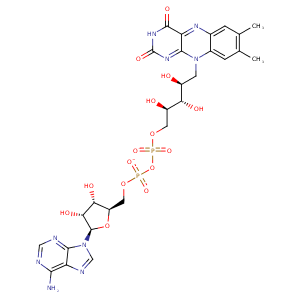
| HET Code: | FAD |
|---|---|
| Formula: | C27H31N9O15P2 |
| Molecular weight: | 783.534 g/mol |
| DrugBank ID: | DB03147 |
| Buried Surface Area: | 76.66 % |
| Polar Surface area: | 381.7 Å2 |
| Number of | |
|---|---|
| H-Bond Acceptors: | 22 |
| H-Bond Donors: | 7 |
| Rings: | 6 |
| Aromatic rings: | 3 |
| Anionic atoms: | 2 |
| Cationic atoms: | 0 |
| Rule of Five Violation: | 3 |
| Rotatable Bonds: | 13 |
| X | Y | Z |
|---|---|---|
| -4.10168 | 7.34842 | -24.1292 |


Represent the protein/ligand binding mode, centered on the ligand
Dashed lines represents hydrogen bonds and metal interactions
Green residue labels for amino acids with hydrophobic contacts (green lines) to the ligand
| Ligand | Protein | Interaction | |||
|---|---|---|---|---|---|
| Atom | Atom | Residue | Distance (Å) | Angle (°) | Type |
| O2A | N | THR- 141 | 3.39 | 164.55 | H-Bond (Protein Donor) |
| C4' | CB | THR- 141 | 4.37 | 0 | Hydrophobic |
| O2P | N | ALA- 142 | 2.79 | 164.98 | H-Bond (Protein Donor) |
| O2B | OE2 | GLU- 164 | 2.66 | 171.52 | H-Bond (Ligand Donor) |
| N3A | N | GLU- 164 | 3.13 | 141.91 | H-Bond (Protein Donor) |
| C1B | CG | GLU- 164 | 3.98 | 0 | Hydrophobic |
| O3B | OD2 | ASP- 165 | 2.72 | 130.61 | H-Bond (Ligand Donor) |
| O2A | CZ | ARG- 172 | 3.79 | 0 | Ionic (Protein Cationic) |
| O3B | NH2 | ARG- 172 | 3.1 | 129.19 | H-Bond (Protein Donor) |
| C8 | CB | ARG- 172 | 3.56 | 0 | Hydrophobic |
| C7M | CB | LEU- 175 | 4.01 | 0 | Hydrophobic |
| C6 | CB | SER- 176 | 4.5 | 0 | Hydrophobic |
| C7M | CB | SER- 176 | 4.14 | 0 | Hydrophobic |
| O4 | NZ | LYS- 177 | 2.86 | 146.37 | H-Bond (Protein Donor) |
| N5 | NZ | LYS- 177 | 3.17 | 134.01 | H-Bond (Protein Donor) |
| N6A | O | VAL- 233 | 2.9 | 154.5 | H-Bond (Ligand Donor) |
| N1A | N | VAL- 233 | 3.05 | 156.79 | H-Bond (Protein Donor) |
| C7M | CE2 | PHE- 284 | 3.83 | 0 | Hydrophobic |
| O2B | NZ | LYS- 286 | 2.84 | 143.66 | H-Bond (Protein Donor) |
| C7M | CD1 | LEU- 311 | 3.79 | 0 | Hydrophobic |
| O3' | OD1 | ASP- 438 | 2.78 | 160.39 | H-Bond (Ligand Donor) |
| C5' | CB | ASP- 438 | 4.43 | 0 | Hydrophobic |
| O1P | N | ASP- 438 | 2.86 | 157.96 | H-Bond (Protein Donor) |
| N1 | N | HIS- 455 | 3.26 | 147.01 | H-Bond (Protein Donor) |
| O2 | N | HIS- 455 | 3.06 | 153.02 | H-Bond (Protein Donor) |
| C2' | CB | HIS- 455 | 4.2 | 0 | Hydrophobic |
| C5' | CB | ALA- 458 | 3.74 | 0 | Hydrophobic |
| O3B | O | HOH- 2013 | 2.7 | 139.54 | H-Bond (Protein Donor) |
| O2P | O | HOH- 2016 | 2.72 | 173.63 | H-Bond (Protein Donor) |
| O2' | O | HOH- 2019 | 2.86 | 162.61 | H-Bond (Ligand Donor) |
| O1P | O | HOH- 2233 | 2.71 | 179.94 | H-Bond (Protein Donor) |
| O1A | O | HOH- 2237 | 2.63 | 179.96 | H-Bond (Protein Donor) |
| O2 | O | HOH- 2521 | 2.7 | 168.88 | H-Bond (Protein Donor) |
