sc-PDB
An Annotated Database of Druggable Binding Sites from the Protein DataBank
An Annotated Database of Druggable Binding Sites from the Protein DataBank
1.900 Å
X-ray
2015-12-30
| Name: | Apoptosis-inducing factor 1, mitochondrial |
|---|---|
| ID: | AIFM1_HUMAN |
| AC: | O95831 |
| Organism: | Homo sapiens |
| Reign: | Eukaryota |
| TaxID: | 9606 |
| EC Number: | 1.1.1 |
| Chain Name: | Percentage of Residues within binding site |
|---|---|
| A | 100 % |
| B-Factor: | 21.872 |
|---|---|
| Number of residues: | 60 |
| Including | |
| Standard Amino Acids: | 54 |
| Non Standard Amino Acids: | 0 |
| Water Molecules: | 6 |
| Cofactors: | |
| Metals: | |
| Ligandability | Volume (Å3) |
|---|---|
| 0.446 | 2038.500 |
| % Hydrophobic | % Polar |
|---|---|
| 41.89 | 58.11 |
| According to VolSite | |
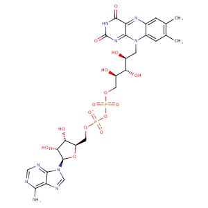
| HET Code: | FAD |
|---|---|
| Formula: | C27H31N9O15P2 |
| Molecular weight: | 783.534 g/mol |
| DrugBank ID: | DB03147 |
| Buried Surface Area: | 77.36 % |
| Polar Surface area: | 381.7 Å2 |
| Number of | |
|---|---|
| H-Bond Acceptors: | 22 |
| H-Bond Donors: | 7 |
| Rings: | 6 |
| Aromatic rings: | 3 |
| Anionic atoms: | 2 |
| Cationic atoms: | 0 |
| Rule of Five Violation: | 3 |
| Rotatable Bonds: | 13 |
| X | Y | Z |
|---|---|---|
| -2.55296 | 11.1847 | -37.1587 |


Represent the protein/ligand binding mode, centered on the ligand
Dashed lines represents hydrogen bonds and metal interactions
Green residue labels for amino acids with hydrophobic contacts (green lines) to the ligand
| Ligand | Protein | Interaction | |||
|---|---|---|---|---|---|
| Atom | Atom | Residue | Distance (Å) | Angle (°) | Type |
| C4' | CB | THR- 141 | 4.43 | 0 | Hydrophobic |
| O2P | N | ALA- 142 | 2.86 | 171.61 | H-Bond (Protein Donor) |
| O2B | OE2 | GLU- 164 | 2.7 | 167.28 | H-Bond (Ligand Donor) |
| N3A | N | GLU- 164 | 3.18 | 148.75 | H-Bond (Protein Donor) |
| C1B | CG | GLU- 164 | 4.04 | 0 | Hydrophobic |
| O2A | NE | ARG- 172 | 3.1 | 159.02 | H-Bond (Protein Donor) |
| O2A | NH2 | ARG- 172 | 3.49 | 138.08 | H-Bond (Protein Donor) |
| O2A | CZ | ARG- 172 | 3.74 | 0 | Ionic (Protein Cationic) |
| C8M | CD | ARG- 172 | 3.69 | 0 | Hydrophobic |
| C8 | CB | ARG- 172 | 3.65 | 0 | Hydrophobic |
| C7M | CB | LEU- 175 | 4.07 | 0 | Hydrophobic |
| C6 | CB | SER- 176 | 4.28 | 0 | Hydrophobic |
| C7M | CB | SER- 176 | 4.15 | 0 | Hydrophobic |
| O4 | NZ | LYS- 177 | 2.61 | 155.9 | H-Bond (Protein Donor) |
| N6A | O | VAL- 233 | 2.95 | 160.87 | H-Bond (Ligand Donor) |
| N1A | N | VAL- 233 | 3 | 157.98 | H-Bond (Protein Donor) |
| C7M | CE1 | PHE- 284 | 3.63 | 0 | Hydrophobic |
| O1A | CZ | ARG- 285 | 3.64 | 0 | Ionic (Protein Cationic) |
| O1A | NH2 | ARG- 285 | 2.74 | 146.15 | H-Bond (Protein Donor) |
| C8M | CG | ARG- 285 | 3.65 | 0 | Hydrophobic |
| O2B | NZ | LYS- 286 | 2.84 | 133.98 | H-Bond (Protein Donor) |
| C7M | CD1 | LEU- 311 | 3.66 | 0 | Hydrophobic |
| O3' | OD1 | ASP- 438 | 2.75 | 161.67 | H-Bond (Ligand Donor) |
| C5' | CB | ASP- 438 | 4.47 | 0 | Hydrophobic |
| O1P | N | ASP- 438 | 2.87 | 156.47 | H-Bond (Protein Donor) |
| N1 | N | HIS- 455 | 3.14 | 162.15 | H-Bond (Protein Donor) |
| O2 | N | HIS- 455 | 3.19 | 138.94 | H-Bond (Protein Donor) |
| C2' | CB | HIS- 455 | 4.12 | 0 | Hydrophobic |
| C5' | CB | ALA- 458 | 3.71 | 0 | Hydrophobic |
| N3 | O | TRP- 483 | 2.94 | 122.46 | H-Bond (Ligand Donor) |
| O2P | O | HOH- 2008 | 2.73 | 165.29 | H-Bond (Protein Donor) |
| O1P | O | HOH- 2137 | 2.68 | 175.99 | H-Bond (Protein Donor) |
| O2 | O | HOH- 2362 | 2.58 | 179.98 | H-Bond (Protein Donor) |
