sc-PDB
An Annotated Database of Druggable Binding Sites from the Protein DataBank
An Annotated Database of Druggable Binding Sites from the Protein DataBank
2.610 Å
X-ray
2015-09-25
| Name: | Ribonuclease 4 |
|---|---|
| ID: | RNAS4_PIG |
| AC: | P15468 |
| Organism: | Sus scrofa |
| Reign: | Eukaryota |
| TaxID: | 9823 |
| EC Number: | 3.1.27 |
| Chain Name: | Percentage of Residues within binding site |
|---|---|
| A | 6 % |
| B | 94 % |
| B-Factor: | 42.040 |
|---|---|
| Number of residues: | 17 |
| Including | |
| Standard Amino Acids: | 17 |
| Non Standard Amino Acids: | 0 |
| Water Molecules: | 0 |
| Cofactors: | |
| Metals: | |
| Ligandability | Volume (Å3) |
|---|---|
| 0.487 | 715.500 |
| % Hydrophobic | % Polar |
|---|---|
| 44.34 | 55.66 |
| According to VolSite | |
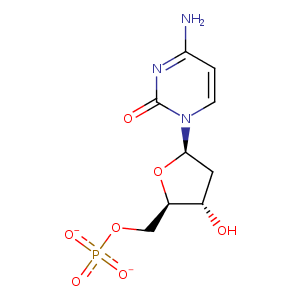
| HET Code: | DCM |
|---|---|
| Formula: | C9H12N3O7P |
| Molecular weight: | 305.181 g/mol |
| DrugBank ID: | - |
| Buried Surface Area: | 38.72 % |
| Polar Surface area: | 170.38 Å2 |
| Number of | |
|---|---|
| H-Bond Acceptors: | 9 |
| H-Bond Donors: | 2 |
| Rings: | 2 |
| Aromatic rings: | 0 |
| Anionic atoms: | 2 |
| Cationic atoms: | 0 |
| Rule of Five Violation: | 0 |
| Rotatable Bonds: | 4 |
| X | Y | Z |
|---|---|---|
| -24.2161 | -31.3873 | -13.8853 |
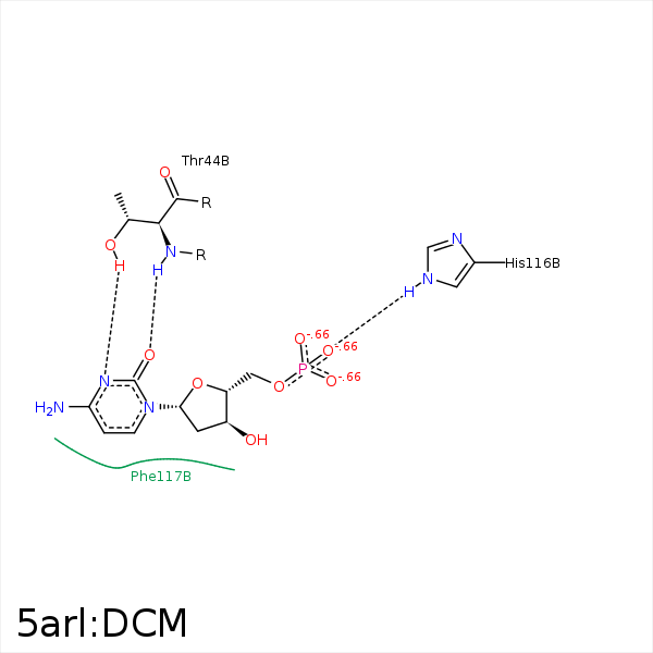


Represent the protein/ligand binding mode, centered on the ligand
Dashed lines represents hydrogen bonds and metal interactions
Green residue labels for amino acids with hydrophobic contacts (green lines) to the ligand
| Ligand | Protein | Interaction | |||
|---|---|---|---|---|---|
| Atom | Atom | Residue | Distance (Å) | Angle (°) | Type |
| O3' | NZ | LYS- 40 | 3.44 | 145.7 | H-Bond (Protein Donor) |
| N3 | OG1 | THR- 44 | 2.99 | 157.43 | H-Bond (Protein Donor) |
| O2 | N | THR- 44 | 2.53 | 125.03 | H-Bond (Protein Donor) |
| O2P | NE2 | HIS- 116 | 2.85 | 127.76 | H-Bond (Protein Donor) |
