sc-PDB
An Annotated Database of Druggable Binding Sites from the Protein DataBank
An Annotated Database of Druggable Binding Sites from the Protein DataBank
2.500 Å
X-ray
2014-12-04
| Name: | DNA ligase |
|---|---|
| ID: | DNLJ_HAEIN |
| AC: | P43813 |
| Organism: | Haemophilus influenzae |
| Reign: | Bacteria |
| TaxID: | 71421 |
| EC Number: | / |
| Chain Name: | Percentage of Residues within binding site |
|---|---|
| A | 100 % |
| B-Factor: | 66.829 |
|---|---|
| Number of residues: | 17 |
| Including | |
| Standard Amino Acids: | 17 |
| Non Standard Amino Acids: | 0 |
| Water Molecules: | 0 |
| Cofactors: | |
| Metals: | |
| Ligandability | Volume (Å3) |
|---|---|
| 0.488 | 411.750 |
| % Hydrophobic | % Polar |
|---|---|
| 59.84 | 40.16 |
| According to VolSite | |
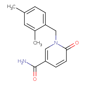
| HET Code: | IWH |
|---|---|
| Formula: | C15H16N2O2 |
| Molecular weight: | 256.300 g/mol |
| DrugBank ID: | - |
| Buried Surface Area: | 53.55 % |
| Polar Surface area: | 63.4 Å2 |
| Number of | |
|---|---|
| H-Bond Acceptors: | 2 |
| H-Bond Donors: | 1 |
| Rings: | 2 |
| Aromatic rings: | 1 |
| Anionic atoms: | 0 |
| Cationic atoms: | 0 |
| Rule of Five Violation: | 0 |
| Rotatable Bonds: | 3 |
| X | Y | Z |
|---|---|---|
| 135.427 | 76.8075 | 25.9235 |
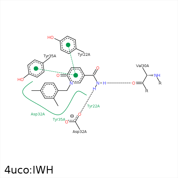


Represent the protein/ligand binding mode, centered on the ligand
Dashed lines represents hydrogen bonds and metal interactions
Green residue labels for amino acids with hydrophobic contacts (green lines) to the ligand
| Ligand | Protein | Interaction | |||
|---|---|---|---|---|---|
| Atom | Atom | Residue | Distance (Å) | Angle (°) | Type |
| C9 | CZ | TYR- 22 | 3.41 | 0 | Hydrophobic |
| N2 | O | VAL- 30 | 3.04 | 164.46 | H-Bond (Ligand Donor) |
| N2 | OD1 | ASP- 32 | 3.06 | 157.71 | H-Bond (Ligand Donor) |
| C9 | CB | ASP- 32 | 4.46 | 0 | Hydrophobic |
| C5 | CB | ASP- 32 | 3.87 | 0 | Hydrophobic |
| C1 | CB | ASP- 36 | 4.11 | 0 | Hydrophobic |
| C3 | CB | ASP- 36 | 4.22 | 0 | Hydrophobic |
