sc-PDB
An Annotated Database of Druggable Binding Sites from the Protein DataBank
An Annotated Database of Druggable Binding Sites from the Protein DataBank
2.910 Å
X-ray
2013-03-04
| Name: | Actin-5C |
|---|---|
| ID: | ACT1_DROME |
| AC: | P10987 |
| Organism: | Drosophila melanogaster |
| Reign: | Eukaryota |
| TaxID: | 7227 |
| EC Number: | / |
| Chain Name: | Percentage of Residues within binding site |
|---|---|
| E | 96 % |
| F | 4 % |
| B-Factor: | 80.721 |
|---|---|
| Number of residues: | 45 |
| Including | |
| Standard Amino Acids: | 44 |
| Non Standard Amino Acids: | 1 |
| Water Molecules: | 0 |
| Cofactors: | |
| Metals: | MG |
| Ligandability | Volume (Å3) |
|---|---|
| 0.810 | 1353.375 |
| % Hydrophobic | % Polar |
|---|---|
| 39.40 | 60.60 |
| According to VolSite | |
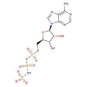
| HET Code: | ANP |
|---|---|
| Formula: | C10H13N6O12P3 |
| Molecular weight: | 502.164 g/mol |
| DrugBank ID: | - |
| Buried Surface Area: | 65.8 % |
| Polar Surface area: | 322.68 Å2 |
| Number of | |
|---|---|
| H-Bond Acceptors: | 16 |
| H-Bond Donors: | 4 |
| Rings: | 3 |
| Aromatic rings: | 2 |
| Anionic atoms: | 4 |
| Cationic atoms: | 0 |
| Rule of Five Violation: | 2 |
| Rotatable Bonds: | 8 |
| X | Y | Z |
|---|---|---|
| 44.7081 | -47.4631 | -30.5858 |
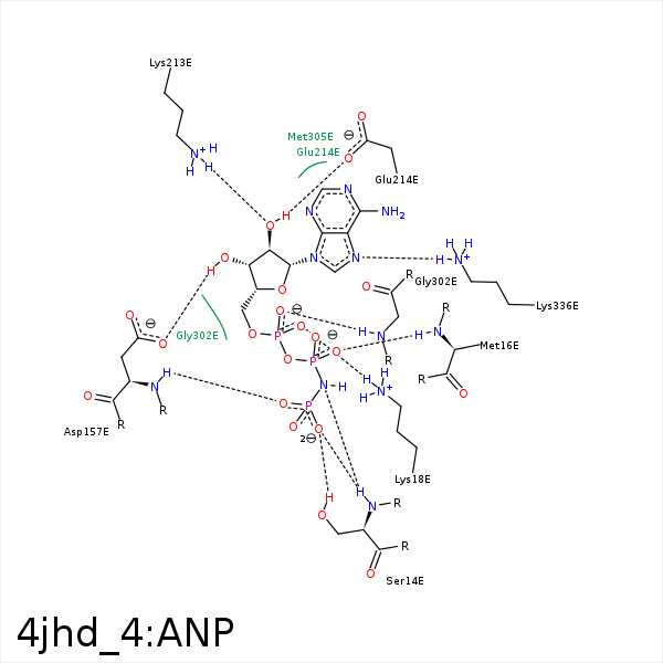


Represent the protein/ligand binding mode, centered on the ligand
Dashed lines represents hydrogen bonds and metal interactions
Green residue labels for amino acids with hydrophobic contacts (green lines) to the ligand
| Ligand | Protein | Interaction | |||
|---|---|---|---|---|---|
| Atom | Atom | Residue | Distance (Å) | Angle (°) | Type |
| O1G | OG | SER- 14 | 3.34 | 132.04 | H-Bond (Protein Donor) |
| O3G | N | SER- 14 | 3.08 | 148.74 | H-Bond (Protein Donor) |
| O3G | OG | SER- 14 | 2.7 | 156.89 | H-Bond (Protein Donor) |
| O2B | N | GLY- 15 | 3.28 | 128.17 | H-Bond (Protein Donor) |
| O2B | N | MET- 16 | 2.94 | 138.65 | H-Bond (Protein Donor) |
| O1B | NZ | LYS- 18 | 3.21 | 0 | Ionic (Protein Cationic) |
| O2B | NZ | LYS- 18 | 3.06 | 0 | Ionic (Protein Cationic) |
| O2A | NZ | LYS- 18 | 3.38 | 0 | Ionic (Protein Cationic) |
| O2A | NZ | LYS- 18 | 3.38 | 175.24 | H-Bond (Protein Donor) |
| O1G | N | ASP- 157 | 2.87 | 146.57 | H-Bond (Protein Donor) |
| C5' | CB | ASP- 157 | 4.35 | 0 | Hydrophobic |
| O3' | OD1 | ASP- 157 | 3.19 | 142.06 | H-Bond (Ligand Donor) |
| O1G | N | GLY- 158 | 3.39 | 155.31 | H-Bond (Protein Donor) |
| O2' | NZ | LYS- 213 | 2.71 | 151.92 | H-Bond (Protein Donor) |
| C2' | CG | GLU- 214 | 4.45 | 0 | Hydrophobic |
| O2' | OE2 | GLU- 214 | 3.13 | 141.24 | H-Bond (Ligand Donor) |
| O1A | N | GLY- 302 | 3.25 | 173.16 | H-Bond (Protein Donor) |
| N7 | NZ | LYS- 336 | 3.21 | 148.62 | H-Bond (Protein Donor) |
| O2G | MG | MG- 402 | 2.58 | 0 | Metal Acceptor |
| O1B | MG | MG- 402 | 1.96 | 0 | Metal Acceptor |
